
ADHD Isn't a Static Brain: What New Long-Term MRI Research Is Revealing
A 2026 systematic review screened 3,385 studies and read the 32 that followed the same children with MRI over years. It found differences that persist, differences that shrink, and one analysis showing no difference at all — and the authors are explicit that this should be read with caution.
- Screened 3,385 studies, included 32 longitudinal MRI studies, baseline ages 4–18 (Ishii-Takahashi et al., Psychiatry and Clinical Neurosciences, 2026).
- Cortical thickness, surface area and gyrification showed a delayed peak in prefrontal, temporal and occipital regions — later, not absent.
- Some differences narrowed with age, particularly in cerebellar white matter and the caudate. Others did not change at all.
- A meta-analysis of change in total grey matter volume found no significant difference between the two groups.
Two things are true about the brains of children with ADHD, and they are usually reported separately because together they are harder to summarise. The first is that differences are measurable, repeatedly, across decades of imaging work. The second is that many of those differences are not fixed — they move with age, and some of them close. A systematic review published in 2026 set out to hold both at once, and the result is a genuinely mixed picture rather than a tidy one.
What the Review Actually Did
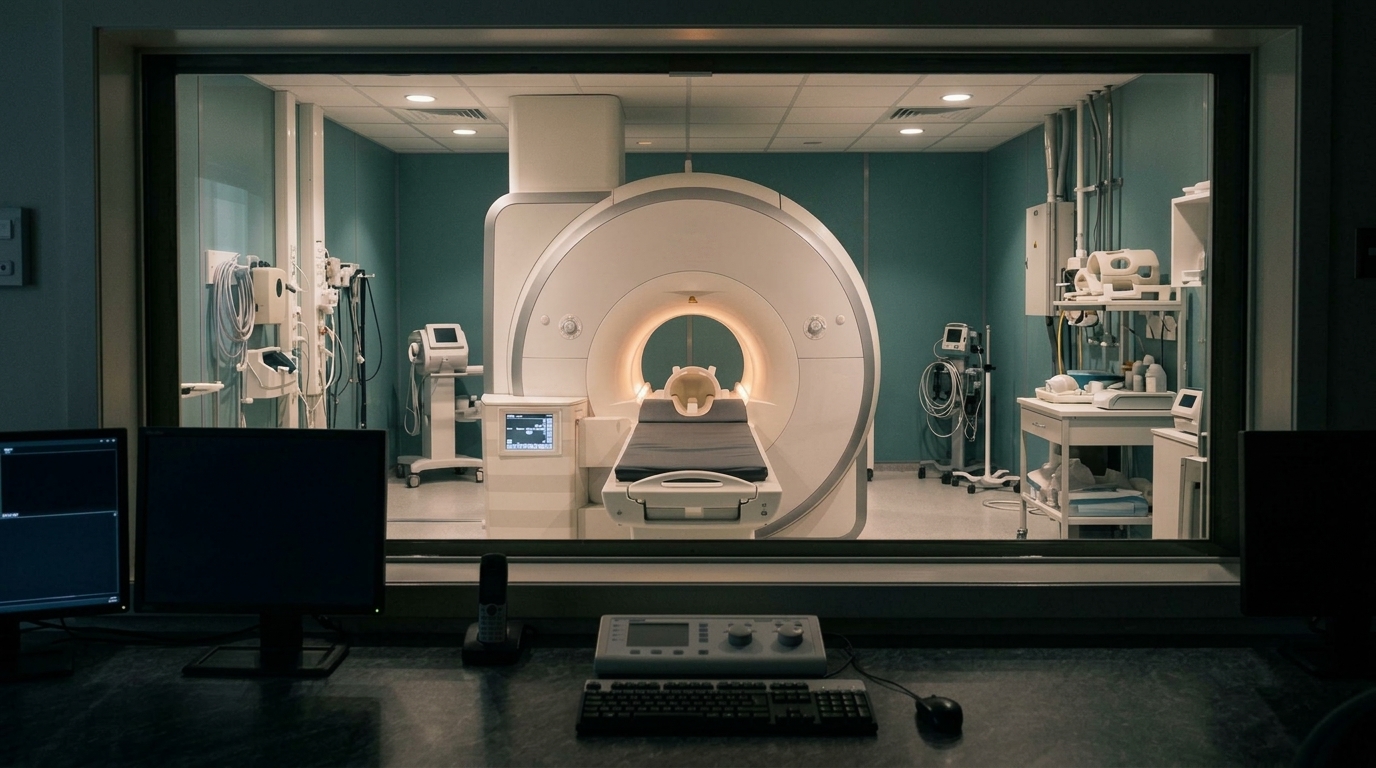
The team screened 3,385 papers published in English up to 9 September 2025 on paediatric ADHD, and selected the 32 that met the criterion that matters here: they followed the same children over time with repeated MRI, rather than comparing different children once. Baseline ages ranged from 4 to 18 (Ishii-Takahashi et al., Psychiatry and Clinical Neurosciences, 2026).
That distinction is the whole value of the exercise. A single scan can tell you that two groups differ today. Only repeated scans of the same children can tell you whether a difference is a delay, a permanent divergence, or something that closes on its own.
The Finding That Suggests a Delay
Cortical thickness, surface area and gyrification showed a delayed peak in children with ADHD, in regions including the prefrontal, temporal and occipital gyri. Delayed, not missing — the developmental curve had the same shape and arrived later.
This is consistent with the finding that made the idea famous. In 2007 a team estimated cortical thickness at more than 40,000 points across 824 MRI scans from 223 children with ADHD and 223 typically developing controls, explicitly to test whether ADHD represents a delay in maturation or a departure from the typical template (Shaw et al., PNAS, 2007). Framing it as a timing difference rather than a deficit was the contribution.
For anyone reading about a child, that framing matters. A curve that arrives late is a different thing from a curve that never arrives.
The Finding That Argues Against a Simple Delay
The same review reports something that does not fit the delay story cleanly. Children with ADHD showed smaller cerebral and cerebellar volumes with no significant change in their developmental trajectories — a difference present at baseline and still present later, tracking in parallel rather than catching up.
It also notes atypical growth patterns that are not slower versions of typical ones, such as contraction of the ventral striatum surface area.
So within one review: some measures look late, some look persistently different, and at least one looks like a different pattern altogether. These are not competing summaries of the same data. They are different measures behaving differently.
And a Third Result That Complicates Both
Two further findings pull the picture in yet another direction. Differences in brain volume and cortical thickness between the two groups diminished with age, particularly in cerebellar white matter and the caudate.
And when the reviewers pooled longitudinal changes in total grey matter volume across studies, the meta-analysis found no significant difference between children with ADHD and their typically developing peers.
A null result at the whole-brain level alongside regional differences is not a contradiction — it is what you would expect if the differences are regional and partly offsetting. But it is a useful corrective to the idea that there is a single global measure on which one group is simply behind.
The Caution the Authors Put in Themselves
The review does not present its own findings as settled. The authors state plainly that because of inconsistencies across studies and the methodological difficulty of integrating them, the results should be interpreted with caution, and they name selection bias as a further concern.
That caveat is doing real work and it is usually the first thing dropped when findings like these are summarised. Longitudinal imaging studies are expensive and hard to retain participants in, they use different scanners, different processing pipelines and different age windows, and the children who enrol and stay are not a random sample of all children with ADHD.
A review that says so is more useful than one that does not — and it is the reason none of the numbers above should be read as a settled fact about brain development.
What This Does and Does Not Tell a Parent
It does not tell you anything about one child. These are group averages assembled across studies, and no finding in this review can be applied to an individual scan, let alone to a child who has never had one. Brain imaging is not used to diagnose ADHD, and nothing here changes that.
What it does support is a narrower and more useful claim: the developmental picture is not static, and several of the measured differences change over the years the studies covered. Whatever is true of a nine-year-old’s measurements is not automatically true of the same child at fifteen.
That is a reason for patience with the timeline rather than optimism about any particular outcome. The related idea — that neural patterns remain modifiable rather than fixed — is covered in what the evidence actually supports about adaptability, and the everyday version of attention demand appears in how thinking changes under pressure.
What the Longitudinal Evidence Supports — and What It Doesn't
Timing, Not Only Deficit
Cortical thickness, surface area and gyrification peaked later rather than failing to develop — a delay in the curve, in specific regions.
Not Everything Catches Up
Smaller cerebral and cerebellar volumes showed no significant change in trajectory. Some differences narrowed; others tracked in parallel.
No Whole-Brain Story
Pooled longitudinal change in total grey matter volume showed no significant group difference. The differences are regional.
Not a Diagnostic Tool
Group averages across 32 studies say nothing about an individual child. Imaging is not used to diagnose ADHD.

Frequently Asked Questions
Does this mean children with ADHD simply catch up?
Some measures appear to. The review found that differences in brain volume and cortical thickness diminished with age, particularly in cerebellar white matter and the caudate. Other measures did not — smaller cerebral and cerebellar volumes showed no significant change in developmental trajectory. Both are in the same review, and treating either as the whole story misreads it.
What does a "delayed peak" in cortical thickness mean?
Cortical thickness rises and then declines across childhood and adolescence. A delayed peak means the curve reached its high point later, in regions including the prefrontal, temporal and occipital gyri, rather than being absent or flat. The idea was established by earlier work estimating thickness at over 40,000 points across 824 scans (Shaw et al., PNAS, 2007).
Can an MRI scan diagnose ADHD?
No. Every finding described here is a group-level average assembled across studies, with substantial variation between individuals inside each group. Diagnosis is clinical, and nothing in this review changes that.
How confident should anyone be in these findings?
Moderately, and the authors say so themselves. They state the results should be interpreted with caution because of inconsistencies across studies and the difficulty of integrating them methodologically, and they name selection bias as a further concern. Longitudinal imaging is hard to do, and the children who enrol and remain are not a random sample.
Why do longitudinal studies matter more than single scans here?
Because only repeated scans of the same children can distinguish a delay from a permanent difference. A single comparison shows that two groups differ now; it cannot show whether that difference is arriving late, closing on its own, or staying put. That is why the review included only the 32 studies that followed the same participants.
Sources
- Ishii-Takahashi A, Yoshimaru Y, Muhammad D, Kawahara T, Yamaguchi R. Longitudinal MRI studies of children with attention-deficit/hyperactivity disorder: a systematic review — Psychiatry and Clinical Neurosciences, July 2026
- Shaw P, Eckstrand K, Sharp W, et al. Attention-deficit/hyperactivity disorder is characterized by a delay in cortical maturation — Proceedings of the National Academy of Sciences, November 2007
Measuring How a Nervous System Is Regulating Now
Research describes groups across years. Understanding one person means measuring that person today. NeuroBalance is a small independent practice in Los Angeles — private one-to-one sessions, the same practitioner each visit, in a quiet setting, over fourteen years. A brain health assessment is where that starts.
Schedule Your Free Consultation
Call us: (424) 625-5445 · Los Angeles, CA
Disclaimer: This content is for informational purposes only and does not constitute medical advice, diagnosis, or treatment. LENS Neurofeedback is not FDA-approved for all conditions mentioned. Please consult with a qualified healthcare provider before beginning any new treatment program.

A balanced brain is a better brain for a happier life.