
The Brain After a Frightening Event: Why Some Memories Fade and Others Won’t Let Go
A frightening event does not simply leave an impression — it recruits specific neural systems to consolidate that memory more durably than ordinary ones. Research in affective neuroscience is mapping the circuit: the amygdala enhances consolidation, the hippocampus encodes context, competing populations of intercalated cells in the amygdala govern a switch between fear and safety states, and the prefrontal cortex regulates whether extinction learning is retained.
- A 2004 review in Annual Review of Neuroscience (McGaugh) established that the amygdala modulates the consolidation of emotionally arousing memories through its projections to the hippocampus and cortex — the mechanism by which frightening events are encoded more durably than neutral ones.
- A 2021 Nature study (Hagihara et al.) found that two competing populations of intercalated cells (ITCs) in the amygdala orchestrate a switch between fear and safety expression. Extinction does not delete the fear memory — it changes which ITC population is currently more active.
- A 2013 Nature Reviews Neuroscience paper (Maren, Phan & Liberzon) described how the hippocampus encodes the context of fear memories, making extinction context-specific. When context shifts, the original fear memory can be retrieved even after successful extinction — a phenomenon called renewal.
- A 2012 Annual Review of Psychology review (Milad & Quirk) found across ten years of research that the ventromedial prefrontal cortex is necessary for long-term retention of extinction memory, with stronger vmPFC activation during extinction predicting better extinction recall the following day in human neuroimaging studies.
In 2021, a team of researchers published a paper in Nature whose title reads almost like a stage direction: “Intercalated amygdala clusters orchestrate a switch in fear state.” The word orchestrate is doing precise work. The paper was not reporting that a brain region responds to fear. It was reporting that competing populations of neurons, in dialogue with each other, determine whether the brain expresses fear or expresses safety — and that the outcome depends on which population is winning at any given moment.
Hagihara and colleagues, working with mice, mapped the function of a set of cells that had been anatomically described for years without their full role being understood: the intercalated cell (ITC) clusters of the amygdala. Using optogenetics — a technique that lets researchers activate or silence specific neurons with light — they showed that two competing ITC populations govern a switch. When one population is more active, fear is suppressed. When the other prevails, fear is expressed. The fear memory itself was not being added or removed. What changed was which cellular population was currently in control.
This is a mechanistically specific answer to a question with real-world weight: after something frightening ends, why do some nervous systems settle and others keep responding as though the threat is ongoing? The experimental model is in mice, and translating the findings to human experience requires care. But the basic architecture of the amygdala and its circuits is conserved across mammals, and the intercalated cell finding sits at the intersection of decades of research on emotional memory. To understand what it means, it helps to start with what the amygdala does to memory in the first place.
How the Amygdala Encodes Emotionally Charged Events
The amygdala is sometimes described as the brain’s alarm center, a shorthand that carries some truth but loses the mechanism. A more accurate framing, based on research going back to James McGaugh’s lab at UC Irvine, is that the amygdala is a modulator of memory consolidation — specifically for memories formed during emotional arousal.
In a 2004 review in Annual Review of Neuroscience, McGaugh summarized decades of work documenting that the amygdala does not store most memories itself. What it does is regulate how well other structures — particularly the hippocampus — consolidate memories after they form. Emotionally arousing events trigger the release of norepinephrine and glucocorticoids. The amygdala responds to these signals and, through its projections to other brain regions, amplifies the consolidation of whatever happened in that moment. The result is that emotionally significant events — particularly threatening ones — are encoded more durably than neutral ones.
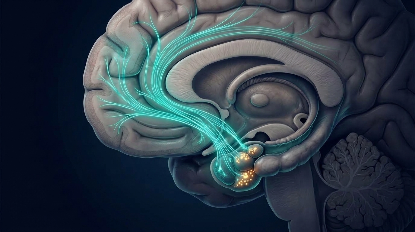
This is not a malfunction. From an evolutionary perspective, preferentially consolidating the details of a dangerous situation makes direct adaptive sense. The organism needs to remember what happened, what signaled it, and where. McGaugh’s review describes this as the amygdala “modulating” consolidation — strengthening the signal that the hippocampus and other regions use to convert short-term experience into long-term memory. The amygdala’s basolateral complex (BLA) projects to the hippocampus and prefrontal cortex; when emotional arousal activates the amygdala, those projections enhance the memory traces being formed elsewhere.
The implication is direct: frightening memories are vivid and persistent because they were consolidated more robustly than the routine moments surrounding them. The research suggests this operates as a gradient. The more emotionally arousing the event, the stronger the consolidation signal. A moderately startling experience is encoded more firmly than a neutral one; an intensely frightening event more firmly still.
The Hippocampus and the Problem of Context
Fear is never learned in a vacuum. An event is frightening in a particular place, at a particular time, with particular cues preceding it. The hippocampus is primarily responsible for binding these contextual elements into the memory alongside the emotional content the amygdala helps consolidate. The result is that fear is not stored as a free-floating association between a cue and a response — it is stored as a contextualized episode.
This has a measurable consequence that Stephen Maren and colleagues described in a 2013 paper in Nature Reviews Neuroscience. The phenomenon is called renewal: when fear is extinguished in one context and then tested in the original context where it was learned, the fear returns. Extinction — the process of learning that a previously threatening signal no longer predicts danger — does not erase the original fear memory. It creates a new, competing memory: a safety memory. The competition between those two memories is context-sensitive. In the original context, the brain tends to retrieve the original fear learning. In the extinction context, it retrieves the safety learning.
Maren’s 2013 review frames this as the “contextual brain” — a model in which the hippocampus does not simply provide spatial background for fear memories but actively governs which memory is retrieved in a given environment. Renewal is one of three well-documented examples of extinction not being permanent: the others are spontaneous recovery (fear returning with the passage of time even without a context shift) and reinstatement (fear returning after exposure to an unrelated stressor). All three reflect the same underlying architecture: extinction creates an inhibitory memory, not an erasure. The hippocampal role in context encoding is central to why that inhibition is conditional.
The Switch: What the 2021 Nature Study Found
The intercalated cells of the amygdala sit between the main input and output regions of the fear circuit. The basolateral amygdala (BLA) receives sensory information and processes associations between cues and outcomes. The central amygdala (CeA) drives the behavioral and physiological outputs of fear — the accelerated heart rate, the freeze response, the hormonal cascade. The ITC clusters sit between these two regions, and their job, as Hagihara and colleagues established, is to regulate the gate.
Using cell-type-specific optogenetics in mice, the team distinguished the functional roles of different ITC populations. They found that two competing clusters inhibit each other: when the extinction-associated population is more active, it suppresses fear expression through the CeA. When the fear-associated network regains the upper hand, it disinhibits the CeA and fear is expressed again. The switch between fear and safety states is not a simple on/off flip of one circuit — it is the outcome of competition between two inhibitory populations, resolved by which is currently more active.
This is the cellular basis of why extinction does not delete the fear memory. The cells encoding the fear association are still present and still capable of driving fear expression. What changes after successful extinction is the balance of activity between the two competing populations. Stress, a shift in context, or the passage of time can all tip that balance back toward fear — not by creating anything new, but by altering which population currently prevails.
The intercalated cell finding also connects upward to the prefrontal cortex. The ventromedial prefrontal cortex (vmPFC) sends projections to the ITC clusters; this is one of the cellular mechanisms through which the prefrontal cortex influences what the amygdala expresses. The competition at the ITC level is not resolved solely by intrinsic amygdala dynamics — it is continuously shaped by descending input from higher cortical regions.
The Prefrontal Cortex and the Architecture of Extinction
If the ITC clusters are the gate, the vmPFC is one of the structures that helps hold it open. Research by Mohammed Milad and Gregory Quirk, summarized in a 2012 review in Annual Review of Psychology covering ten years of progress in fear extinction research, established the prefrontal contribution to extinction memory through both animal and human studies.
In rodents, the infralimbic cortex — the region considered broadly homologous to the human vmPFC — is necessary for the long-term retention of extinction memory. When infralimbic activity is suppressed after extinction training, animals can acquire extinction normally but cannot recall it the following day: the safety learning is lost overnight. Conversely, the prelimbic cortex, which projects to the BLA and promotes fear expression, opposes the infralimbic. Two competing prefrontal regions, at a higher level of the circuit, recapitulate the same logic as the competing ITC populations: the output depends on which side of the competition is more active.
In humans, Milad and Quirk’s 2012 review described neuroimaging studies finding that participants who show stronger vmPFC activation during extinction learning show better extinction memory tested the following day. Participants with reduced vmPFC thickness or activity — a pattern found in neuroimaging studies of individuals experiencing symptoms associated with traumatic stress — also show reduced extinction recall. The vmPFC contribution is not to the initial formation of the extinction memory but to its consolidation and later retrieval.
The clinical implication is not straightforward, and Milad and Quirk’s review is careful about this. The vmPFC finding describes a population-level pattern. It does not explain what drives reduced vmPFC engagement in any individual, or whether that pattern is a cause or consequence of persistent fear responding. Those questions remain open in the current literature.
What the Evidence Does Not Tell Us
The circuit-level picture of fear memory — amygdala consolidation, hippocampal context encoding, ITC competition, prefrontal extinction regulation — has advanced substantially over the past two decades. But several limits apply to what the current evidence can support.
Most of the mechanistic work reviewed here was conducted in rodents, using tools — particularly optogenetics — that are not available in human studies. In a 2024 review of the circuits for adaptive regulation of fear and extinction memory, Plas, Tuna, and colleagues (including Maren, as last author) summarized where the translation from rodent to human is strong and where it remains uncertain. The basic architecture of the amygdala-hippocampus-vmPFC circuit is broadly conserved across mammals, but the human amygdala is more anatomically complex than the mouse amygdala, and the relative contribution of different components has not been measured in living human tissue with the cellular resolution that optogenetics provides. Human studies rely primarily on neuroimaging, which measures correlates of neural activity at a spatial resolution that cannot distinguish adjacent cell types.
The research also describes population-level patterns, not individual predictions. A study finding that higher vmPFC activation correlates with better extinction recall describes a relationship across a sample of participants. It does not predict, for any individual, how their extinction learning will proceed. Individual differences in fear learning and extinction are substantial — far more substantial than circuit models suggest. The sources of that variation include genetics, developmental history, prior experience, and current physiological state. A circuit model identifies architecture; it does not capture the range of what people actually experience, and the 2024 Plas et al. review acknowledges this gap directly as an area of ongoing investigation rather than a settled question.
Four Circuit Components That Influence How Fear Memories Are Held and Regulated
Amygdala consolidation
The basolateral amygdala enhances memory consolidation during emotional arousal through its projections to the hippocampus and cortex. This is the mechanism by which frightening events tend to be remembered more vividly than neutral ones (McGaugh, Annual Review of Neuroscience, 2004).
Hippocampal context encoding
The hippocampus binds contextual details — where and when — into fear memories. Extinction learning is context-specific: the original fear memory remains accessible in the original context, producing the renewal effect (Maren, Phan & Liberzon, Nature Reviews Neuroscience, 2013).
Intercalated cell competition
Two competing populations of intercalated cells (ITCs) in the amygdala act as a regulatory gate between fear and safety states. Extinction changes which population is currently dominant, without deleting the cells encoding fear (Hagihara et al., Nature, 2021).
Prefrontal extinction regulation
The vmPFC projects to the ITC clusters and is necessary for retaining extinction memory overnight. Stronger vmPFC activation during extinction predicts better extinction recall the following day in human neuroimaging studies (Milad & Quirk, Annual Review of Psychology, 2012).
Frequently Asked Questions
Why are memories of frightening events often more vivid than memories of ordinary moments?
Because frightening events activate the amygdala, which in turn enhances how other brain regions consolidate the memory. A 2004 review by McGaugh in Annual Review of Neuroscience described this as the amygdala modulating consolidation through stress-hormone signaling, including norepinephrine and glucocorticoids. The result is a memory that is encoded more robustly than a neutral event from the same period. This is not a pathological process — it reflects an architecture that preferentially retains information about dangerous situations.
What does it mean that the brain has a ‘fear switch’ rather than simply deleting fear memories?
Research by Hagihara and colleagues (Nature, 2021) found that two competing populations of intercalated cells in the amygdala govern whether fear is expressed or suppressed. Extinction learning — learning that a once-threatening signal is now safe — does not remove the cells encoding the original fear association. It changes the balance of activity between the two competing populations. Because the fear-encoding cells remain present and functional, the original response can return when context shifts, stress occurs, or time passes. This architecture explains why extinction is not permanent erasure.
Why does fear sometimes return even after it seemed to go away?
Because extinction creates an inhibitory memory that competes with the original fear memory rather than replacing it, and that competition is context-sensitive. A 2013 paper in Nature Reviews Neuroscience by Maren, Phan, and Liberzon described three documented forms of this return: renewal (fear returning in the original context), spontaneous recovery (fear returning with the passage of time), and reinstatement (fear returning after an unrelated stressor). All three reflect the hippocampus’s role in context-dependent retrieval: which memory is expressed depends on the current environment and the relative activation of competing neural populations.
What role does the prefrontal cortex play in managing responses to frightening memories?
The ventromedial prefrontal cortex (vmPFC) — and its rodent analog, the infralimbic cortex — is necessary for retaining extinction memory overnight. Research reviewed by Milad and Quirk in a 2012 Annual Review of Psychology paper found that animals with suppressed infralimbic activity can acquire extinction normally but cannot recall it the next day. In human neuroimaging studies, stronger vmPFC activation during extinction learning predicts better extinction recall tested later. The prefrontal contribution is to consolidation and retrieval of safety learning, not to the initial formation of that learning.
What are the main limits on what the current research can tell us?
Most mechanistic work on the fear circuit has been conducted in rodents using optogenetics, a tool not available in human research. Human studies rely on neuroimaging, which operates at a spatial resolution that cannot distinguish individual cell types. A 2024 review by Plas, Tuna, and colleagues in Frontiers in Behavioral Neuroscience acknowledged that while the basic amygdala-hippocampus-vmPFC architecture is broadly conserved across mammals, direct comparisons carry real uncertainty. The research also describes population-level patterns — group averages across samples of participants — not individual predictions. Individual differences in fear learning and extinction are substantial, and their sources are not fully accounted for by current circuit models.
Sources
- The amygdala modulates the consolidation of memories of emotionally arousing experiences — McGaugh JL — Annual Review of Neuroscience, 2004
- Intercalated amygdala clusters orchestrate a switch in fear state — Hagihara KM et al. — Nature, 2021
- The contextual brain: implications for fear conditioning, extinction and psychopathology — Maren S, Phan KL, Liberzon I — Nature Reviews Neuroscience, 2013
- Fear extinction as a model for translational neuroscience: ten years of progress — Milad MR, Quirk GJ — Annual Review of Psychology, 2012
- Neural circuits for the adaptive regulation of fear and extinction memory — Plas SL, Tuna T, Bayer H et al. — Frontiers in Behavioral Neuroscience, 2024
Questions About Nervous System Regulation and Brain Health
If you have questions about how the brain responds to stress and emotional experience, we offer a free initial consultation. We work with both children and adults in a private, one-to-one setting — the same practitioner, the same room, every session. Learn about what to expect.
Schedule Your Free Consultation
Call us: (424) 625-5445 · Los Angeles, CA
Disclaimer: This content is for informational purposes only and does not constitute medical advice, diagnosis, or treatment. LENS Neurofeedback is not FDA-approved for all conditions mentioned. Please consult with a qualified healthcare provider before beginning any new treatment program.

A balanced brain is a better brain for a happier life.