
Sleep, Stress and Alzheimer’s: A Three-Way Conversation Inside the Brain
Research on the brain’s nightly clearance system and the stress-hormone pathway is raising specific biological questions about whether sleep quality and chronic stress may be modifiable contributors to Alzheimer’s disease — and how the evidence currently stands.
Alzheimer’s disease is widely thought of as something that happens with age — written in genes, sealed by biology, accumulating through processes that begin long before any symptom appears. The advice that follows from this view tends toward early detection and planning. Prevention, if mentioned at all, is presented as speculative at best.
That framing is not entirely wrong. Genetic factors do influence risk, and pathological changes in the brain can precede symptoms by decades. But it is a partial account of the current science, and the part it leaves out has been getting considerably more attention in recent years.
Four lines of research — on the brain’s nightly waste-clearance system, on the mechanics of how that system works, on how chronic stress alters brain chemistry, and on a small region of the brainstem where these processes intersect — are beginning to put two specific questions into sharper focus: whether sleep quality matters to Alzheimer’s pathology, and whether chronic stress contributes to it. Neither question has a clean answer. Both are worth understanding carefully.
The Brain Has a Nightly Cleaning Cycle

During NREM slow-wave sleep — the deeper, restorative stages that most people reach within the first few hours of sleep — a clearance system distributed throughout the brain becomes significantly more active. This system, called the glymphatic system, uses channels built into the walls of small blood vessels to move cerebrospinal fluid through brain tissue. It works somewhat like an irrigation channel: fluid enters through one set of channels, flows through the narrow interstitial spaces between neurons, and exits through a second set, carrying metabolic waste products alongside it.
Among the substances the glymphatic system clears are amyloid-beta (Aβ) and tau — two proteins whose abnormal accumulation is the defining pathological feature of Alzheimer’s disease. These proteins are produced continuously in normal brain activity; the question is how efficiently they are removed.
A 2025 review in Frontiers in Neurology by Corbali and Levey examined the evidence on glymphatic function across neurological conditions. It found that clearance relies critically on proteins called aquaporin-4 (AQP4) channels, expressed densely in the endfeet of astrocytic cells lining the vessel walls. When these channels are disrupted, glymphatic flow slows. The review also noted that a measure of glymphatic efficiency derived from diffusion-weighted imaging — the DTI-ALPS index — shows reduced values in people with conditions associated with Aβ accumulation. Lower DTI-ALPS scores tracked with higher amyloid burden in multiple studies the authors examined (Corbali and Levey, Frontiers in Neurology, 2025).
What makes this relevant to sleep specifically: measurements in animal models found that the interstitial space — the gap through which fluid has to flow — expands by approximately 60% during slow-wave sleep compared to waking. NREM Stage 3, the deepest sleep stage, appears to be when clearance peaks.
How Scientists Traced the Clearance Signal
Understanding that glymphatic clearance peaks during slow-wave sleep raised a further question: what drives the increased activity? A 2025 study published in Cell by Hauglund and colleagues identified a specific mechanism: slow oscillations in the brain’s concentration of norepinephrine during NREM sleep.
Norepinephrine is a neurotransmitter most commonly associated with attention and arousal. During NREM sleep, the brain’s principal norepinephrine source — a small region in the brainstem called the locus coeruleus — maintains a rhythm rather than switching off. Norepinephrine concentration rises and falls in slow cycles, approximately every 50 seconds. These oscillations, the researchers found, are coupled to tiny rhythmic contractions of blood vessels — called slow vasomotion — that function like a pump, pulsing cerebrospinal fluid through the surrounding tissue.
To test whether the oscillations caused the flow or merely accompanied it, the researchers used the sleep medication zolpidem, which suppresses deep sleep architecture. Zolpidem reduced norepinephrine oscillations by approximately 50%. Glymphatic transport — measured by tracking where a tracer moved through the brain during sleep — dropped by more than 30% (Hauglund et al., Cell, 2025).
This is worth noting carefully. The study was conducted in rodent models. It identified a mechanism plausibly linked to clearance; it did not follow Alzheimer’s pathology as the endpoint. What it establishes is a credible biological pathway connecting sleep architecture to the efficiency of Aβ removal — not a human clinical result on Alzheimer’s disease itself.
What Chronic Stress Does to the Same Chemistry
The second line of research converges from a different direction. Chronic psychological stress activates the hypothalamic-pituitary-adrenal (HPA) axis — the body’s primary stress-response system — producing sustained elevation of the hormone cortisol.
A 2024 review in Cureus by Sharan and Vellapandian examined the evidence on HPA axis dysregulation and Alzheimer’s-related pathology. It documented three converging observations from the existing literature: elevated cortisol is associated with increased production of amyloid-beta; chronic stress conditions are associated with hyperphosphorylation of the tau protein, a process that promotes its aggregation into the tangles associated with neurodegeneration; and the comorbidity rate between depression — which frequently involves sustained HPA axis activation — and Alzheimer’s disease is estimated at 20 to 30% in clinical populations. The authors note that the directionality of the depression-Alzheimer relationship is not fully established: whether depression contributes to neurodegeneration, whether early-stage neurodegeneration creates conditions for depression, or whether the two share common upstream factors remains an open question (Sharan and Vellapandian, Cureus, 2024).
There is a second, indirect route through which chronic stress likely affects Alzheimer’s-related pathology: it disrupts sleep. Sustained cortisol elevation is associated with difficulty entering and maintaining slow-wave sleep — the stage during which glymphatic clearance is most active. This creates a potential compounding effect: stress degrades sleep architecture, and degraded sleep architecture reduces the nightly clearance of the proteins whose accumulation defines Alzheimer’s pathology. Whether this compounding is clinically significant in humans has not been established, but the biological reasoning is internally consistent.
The Locus Coeruleus: Where the Threads Meet
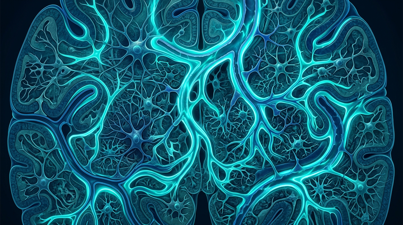
The locus coeruleus sits at the intersection of these two pathways in a way that is not incidental. It is the brain’s primary source of norepinephrine — both the signal that drives glymphatic clearance during sleep and a key modulator of the stress response when the HPA axis activates. The same structure is responsible for both functions.
It is also one of the earliest sites of tau accumulation in the Alzheimer’s disease process. Neuropathological studies have documented tau deposits in the locus coeruleus before they appear in the hippocampus or cortex — sometimes decades before symptoms. This matters for both threads: if early tau pathology in the locus coeruleus degrades the quality of its norepinephrine output, that degradation could reduce the oscillation-driven vasomotion that Hauglund and colleagues identified as the clearance mechanism during NREM sleep. And if chronic stress hyperstimulates the locus coeruleus over years, that overstimulation may contribute to the oxidative stress and neuroinflammatory conditions associated with tau accumulation in the region.
The locus coeruleus appears, in short, to be both a biological hub and a candidate for the mechanism by which sleep disruption and chronic stress might compound each other — and through which both might connect to Alzheimer’s-related pathology. Whether this sequence plays out in humans the way animal models suggest is the question the field is now designing studies to answer.
What the Evidence Does and Does Not Establish
A 2026 systematic review in the Journal of Clinical Medicine by Cabarkapa and colleagues examined the sleep-glymphatic-Alzheimer’s connection directly. Pooling data from four eligible studies involving 397 participants, the review found that better slow-wave sleep architecture was associated with better glymphatic function, as measured by the DTI-ALPS index. Participants with more fragmented sleep or less time in deep NREM showed lower glymphatic efficiency scores.
But the authors were explicit about the limitations. All four studies in the review were cross-sectional — they measured sleep and glymphatic function at a single point in time, in the same individuals, without following them forward. A cross-sectional design can document an association, but it cannot establish which came first. It cannot determine whether poor sleep reduces glymphatic function, whether early-stage neurodegeneration disrupts both simultaneously, or whether a third variable influences both independently (Cabarkapa et al., Journal of Clinical Medicine, 2026).
The literature as a whole faces this constraint. No randomized controlled trial has demonstrated that improving sleep in healthy adults reduces Alzheimer’s risk. No long-term human intervention has shown that reducing chronic stress measurably alters the trajectory of Alzheimer’s pathology. What exists are biologically coherent mechanisms, epidemiological associations, and a consistent pattern in animal models — a scientific foundation for the research questions, not final answers to them.
The Hauglund 2025 findings were in rodents. The Corbali and Levey 2025 review drew on studies with modest sample sizes. Group-level statistics describe populations; they do not say what any individual’s sleep history or stress exposure means for their own brain. These are honest limits, worth stating plainly, because the questions being asked are important enough to warrant rigorous investigation rather than premature conclusions in either direction.
Frequently Asked Questions
What is the glymphatic system?
The glymphatic system is a network of channels built into the walls of small blood vessels in the brain, through which cerebrospinal fluid moves to clear metabolic waste — including amyloid-beta and tau — from brain tissue. The system is substantially more active during deep NREM slow-wave sleep, which is the period when most of this clearance is believed to occur.
Does poor sleep cause Alzheimer’s disease?
The current evidence does not establish a direct causal relationship. What it shows is an association: people with more fragmented sleep or less time in slow-wave sleep tend to show lower glymphatic efficiency scores, and lower efficiency has been linked to higher amyloid burden. The studies are largely cross-sectional, so they cannot determine whether disrupted sleep reduces clearance, whether early neurodegeneration disrupts sleep, or both. Causality has not been established in human trials.
Can chronic stress contribute to Alzheimer’s risk?
Research has documented associations: elevated cortisol with increased amyloid-beta production, chronic stress with tau hyperphosphorylation, and a 20–30% comorbidity rate between depression and Alzheimer’s disease. Whether the relationship is causal, and in which direction, has not been determined. Chronic stress also disrupts slow-wave sleep, which may compound its effects through the glymphatic pathway.
What is the locus coeruleus, and why does it appear in this research?
The locus coeruleus is a small region in the brainstem that produces most of the brain’s norepinephrine. It is relevant for two reasons: its norepinephrine oscillations during NREM sleep appear to drive the vasomotion that powers glymphatic clearance, and it is one of the first sites in the brain where tau deposits appear in Alzheimer’s disease — sometimes decades before symptoms. Both the sleep-clearance pathway and early Alzheimer’s vulnerability pass through this same region.
What does this research mean for daily choices about sleep and stress?
The research identifies biologically plausible mechanisms, not clinical recommendations. It does not establish that improving sleep reduces an individual’s Alzheimer’s risk, and translating group-level associations into personal guidance requires a conversation about individual circumstances. What it offers is a clearer picture of why sleep quality and chronic stress are considered worth studying in this context — and a scientific basis for the ongoing research designed to answer the causal question more directly.
Sources
- Corbali O and Levey AI (2025). The glymphatic system and its role in neurological disorders. Frontiers in Neurology, 16:1543725. — Frontiers in Neurology
- Hauglund NL et al. (2025). Norepinephrine-mediated slow vasomotion drives glymphatic clearance during sleep. Cell, 189(1), 100-116. — Cell (Elsevier)
- Cabarkapa S et al. (2026). Sleep architecture, glymphatic function, and Alzheimer's disease pathology: a systematic review. Journal of Clinical Medicine, 15(7), 2463. — Journal of Clinical Medicine (MDPI / PMC)
- Sharan B and Vellapandian C (2024). The role of the HPA axis in Alzheimer's disease. Cureus, 16(9), e67595. — Cureus
Understand Your Nervous System
If you have been experiencing symptoms associated with poor sleep, difficulty managing stress, or changes in concentration or recall, a brain health assessment can help clarify where your nervous system is currently functioning and identify areas that may benefit from support.
Schedule Your Free Consultation
Call us: (424) 625-5445 · Los Angeles, CA
Disclaimer: This content is for informational purposes only and does not constitute medical advice, diagnosis, or treatment. LENS Neurofeedback is not FDA-approved for all conditions mentioned. Please consult with a qualified healthcare provider before beginning any new treatment program.

A balanced brain is a better brain for a happier life