
The Brain's 'Switching System': Why Moving From One Task to Another Can Be So Hard
A look at what neuroimaging research shows about how the brain coordinates task-switching — and why equivalent performance on switch-cost tests can coexist with substantially different patterns of neural engagement.
Two findings from the same decade of neuroimaging research pull in opposite directions, and the space between them is where the real question lives.
The first: many people who report that switching between tasks is genuinely hard — who lose their footing when interrupted, who need a beat of quiet to re-enter what they were doing, who find transitions disproportionately costly regardless of what the tasks are — often do not score worse than their peers on clinical task-switching tests. Their switch costs, measured in reaction time and accuracy, frequently fall within normal ranges. By the numbers, the difficulty appears not to be there.
The second: when researchers watch what happens in those same brains during the switch — using neuroimaging to track which regions activate and which route the nervous system takes — the picture changes substantially. The brain arrives at the same behavioral answer by a different path. Different circuits engage. Different regions compensate. And the path taken may have a different cost structure than the one it is being compared against.
The gap between those two findings is not a contradiction to be resolved by picking a side. It is the finding. Understanding what it means requires tracing what task-switching actually asks of the nervous system, and why the asking is more complicated than any single region or any single explanation.
The Signal That Starts the Switch

Task-switching research has long focused on documenting where switching is coordinated and how long it takes. A 2024 study by Cole and colleagues, published in Nature Communications, sharpened the question considerably: instead of asking which brain regions are associated with task-switching, they asked which are causally necessary for it to happen quickly.
Their answer centered on the anterior cingulate cortex, a structure running along the medial wall of the frontal lobe, positioned where attention monitoring, error detection, and the adjustment of cognitive strategy converge. Cole et al. (Nature Communications, 2024) found that the ACC generates prediction-error signals — responses to the mismatch between what the brain expected to happen next and what actually happened — and that these signals are required for rapid task-switching to occur. When the ACC’s prediction-error output was disrupted experimentally, switching slowed.
The mechanism involved a disinhibitory interneuron circuit: a configuration in which one type of inhibitory cell releases a second type from suppression, allowing a new signal to propagate quickly. The ACC does not simply detect that something has changed; it actively dislodges the brain’s current task representation so that a new one can take hold.
Cole et al. also found that what they called baseline task inertia — how persistently the brain maintains its current context rather than releasing it — predicted individual differences in switch cost magnitude. This is not a finding about effort or motivation. It is about a specific signaling mechanism: how readily the ACC can interrupt the current pattern and authorize the shift. People whose task representations are more resistant to that interruption show larger switch costs, even when their overall cognitive function is otherwise comparable.
The Structural Foundation: White Matter and the Fronto-Striatal Circuit
The ACC prediction-error signal does not operate alone. It participates in a broader circuit that includes the basal ganglia — a set of subcortical structures involved in selecting, maintaining, and updating action programs — and the thalamus, which relays signals between cortex and the rest of the brain. This fronto-striato-thalamic loop is implicated in a wide range of functions requiring flexible updating, and its capacity for supporting cognitive flexibility appears to have a structural component.
A 2014 study by van Schouwenburg and colleagues, published in Neuropsychologia, examined the white matter microstructure of basal ganglia tracts in adults, including individuals who did and did not experience symptoms associated with attention and focus difficulty. White matter is the brain’s connective tissue — myelinated axon bundles whose integrity influences how efficiently signals travel between regions. Van Schouwenburg et al. measured microstructure using diffusion tensor imaging, which tracks the directional movement of water molecules through tissue as a proxy for how well organized the fibers are.
They found that white matter integrity in basal ganglia tracts predicted attention-switching performance across participants — not only in the group with attention-related symptoms, but as a continuous relationship extending across the sample. Individuals whose basal ganglia white matter showed lower organizational integrity tended to show larger switch costs. The fronto-striato-thalamic circuit’s efficiency, as reflected in its structural coherence, predicted how fluidly a person could redirect attention (van Schouwenburg et al., Neuropsychologia, 2014).
A structural finding does not predict any individual’s experience. White matter microstructure is shaped by genetics, development, and environment, and its relationship to any particular behavioral outcome involves many other factors. What the finding establishes is that the circuit’s capacity is not simply a matter of applying more effort in any given moment — it reflects the physical organization of the pathway the signal travels.
The Same Score, a Different Route
The behavioral equivalence paradox appears most directly in studies that compare both performance and brain activation in people who do and do not experience symptoms associated with attention difficulty — and find that equivalent performance masks non-equivalent neural processes.
A 2010 study by Dibbets and colleagues, published in Neuropsychology, placed adult participants with and without ADHD-associated symptoms inside an fMRI scanner during a task-switching paradigm. The behavioral data — reaction times, error rates, switch costs — showed no significant group differences in executive control. Both groups switched comparably by the numbers.
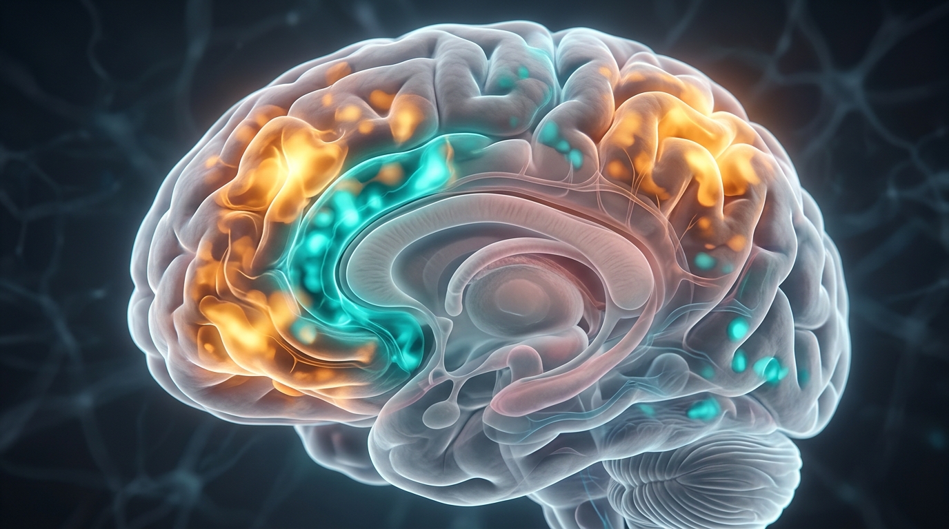
The imaging data separated them. Control participants showed stronger activation in the putamen, medial frontal gyrus, and thalamus during switching — the core fronto-striatal path, consistent with the circuit van Schouwenburg and colleagues associated with structural efficiency. Participants experiencing symptoms associated with attention difficulty showed greater activation in the dorsal anterior cingulate cortex, middle temporal gyrus, and precuneus — regions associated with cognitive monitoring, effort regulation, and self-referential processing, but not the primary route for efficient action selection (Dibbets et al., Neuropsychology, 2010).
The interpretation Dibbets and colleagues offered was that the ADHD group appeared to engage a higher-cortical compensatory route — achieving the required behavioral result through circuits that perform the job less automatically, requiring more top-down monitoring to complete what the fronto-striatal path handles more efficiently in the control group.
The study was not large by current neuroimaging standards, and the interpretation involves inference beyond what fMRI alone can establish: observing greater ACC and precuneus activation is not the same as directly measuring effortfulness or cognitive load. What the study documents directly is a dissociation between behavior and neural routing. The score looks the same; the brain does not.
Across Diagnoses, a Shared Signature of Elevated Effort
A finding in one diagnostic group could reflect something specific to that group. A finding that replicates across multiple very different conditions begins to suggest something more general about how the brain responds when cognitive flexibility is challenged.
A 2025 coordinate-based meta-analysis by Meda and colleagues, published in the European Journal of Neuropsychopharmacology, examined neuroimaging studies of cognitive flexibility across three diagnostic categories: schizophrenia, autism spectrum disorder, and generalized anxiety disorder. These three conditions share little in terms of presentation, etiology, or the populations they affect most heavily. What they share, in a growing imaging literature, is an association with difficulties in task-switching and set-shifting.
Meda et al. screened 22 neuroimaging studies and identified a convergent pattern of hyperactivation — not reduced activation, but elevated activation relative to comparison groups — in what they described as the frontoparietal network. The specific regions: the right medial frontal cortex and anterior cingulate cortex, the right superior parietal lobule, and the left superior temporal gyrus (Meda et al., European Neuropsychopharmacology, 2025).
The pattern held across all three diagnostic groups. This does not mean the three conditions are neurologically identical, or that the same mechanism produces the shared finding in each case. It means that when researchers have observed neural correlates of impaired cognitive flexibility across these populations, they have tended to find the same network working harder than it does in comparison samples — a pattern of extra engagement, not simple failure.
A coordinate-based meta-analysis aggregates peak activation coordinates from many studies; it cannot account for all the methodological variation across them. The 22-study sample is meaningful for this literature but modest in absolute terms. The finding is convergence across diverse conditions — a signal worth noting, not a settled answer.
When Memory Takes Over the Switching Function
For children experiencing symptoms associated with autism spectrum conditions, the deviation from the expected switching network appears to take a specific form — not greater recruitment of cortical monitoring regions, but greater recruitment of memory systems.
A 2020 study by Dirks and colleagues, published in Autism Research, compared neural responses during a set-shifting task in children with ASD and typically developing comparison children. Behavioral performance was again comparable across groups; the divergence was in where the brain recruited resources.
The region showing significantly greater activation in the ASD group was the parahippocampal gyrus — a structure in the medial temporal lobe associated with encoding and retrieving context, processing spatial information, and recognizing familiar environments. It sits at the edge of the hippocampal memory system rather than the frontal-parietal control network that anchors typical task-switching.
Dirks et al. (Autism Research, 2020) interpreted this as evidence that children with ASD may draw on memory-based processing more heavily during set-shifting — recalling prior contexts or strategies associated with the new task rule, rather than generating a prospective update through the frontal control network in the moment of the switch. If the frontal updating mechanism is less readily engaged, a memory-based route may perform a similar function at the cost of depending on prior experience and recognition rather than flexible in-the-moment generation.
The study used a relatively small sample, as is common in pediatric neuroimaging research, and described its paradigm as a “putative” set-shifting task — meaning it was designed to engage set-shifting but its neural profile in this specific population had not been independently validated before publication. The finding is consistent with a broader pattern of atypical cognitive flexibility responses in ASD; the specific mechanism requires further study.
What the Research Does Not Establish
Five studies across two decades point in a consistent direction: the behavioral cost of task-switching does not capture the full picture of how different the underlying process can be. Consistent direction is meaningful. So are the limits of what is established.
All five studies describe group-level comparisons. A group pattern does not translate into a prediction about any person. The individual who experiences every task transition as genuinely costly might show these activation patterns; they might not. The individual who shows elevated frontoparietal activation in a scanner might report no unusual difficulty with transitions in daily life. Group statistics describe tendencies in populations; they do not characterize individuals.
Neuroimaging sample sizes in this literature are typically small. The Dibbets et al. study examined a limited cohort of adults in a single laboratory session. The Dirks et al. study examined children at one site. Even the Meda et al. meta-analysis, which aggregates 22 studies, works with source material that carries those constraints. Larger, multisite, longitudinal studies would substantially strengthen or complicate these conclusions.
The relationship between differential neural activation and the subjective experience of difficulty has not been directly demonstrated in any of these studies. Observing greater ACC or precuneus activation during switching is not the same as measuring experienced effort and finding it elevated. The inference is intuitive — regions associated with cognitive monitoring working harder suggests more monitoring is required — but it is not established by imaging data alone.
The gap between behavioral equivalence and neural difference can be explained by genuine compensation, by ceiling effects in the task design, by a fundamentally different but equally efficient neural strategy, or by some combination. None of these is ruled out. The task-switching paradigms used in laboratory settings may also not capture what makes real-world transitions difficult: switching under fatigue, transitioning under emotional load, re-entering a complex context after an unexpected interruption. The lab task and the lived experience are not the same measurement.
Frequently Asked Questions
What is a task-switching cost and why does the brain pay it?
A task-switching cost is the measurable increase in reaction time and errors that occurs when a person transitions between two different tasks, compared to repeating the same task. The cost exists because the brain must suppress the current task’s rules and activate a new set — and that process takes time. Research by Cole and colleagues, published in Nature Communications in 2024, identified a specific mechanism: prediction-error signals generated in the anterior cingulate cortex are causally required for rapid switching to occur. When the brain’s representation of the current task persists longer than it should — what the researchers called baseline task inertia — the switch cost increases. This cost is a normal feature of cognition; it becomes notable when the magnitude is unusually large or the everyday experience of switching is disproportionate to what task tests capture.
Why might transitions feel hard even when performance tests look normal?
Neuroimaging research suggests that equivalent behavioral performance on a task-switching test does not guarantee that the same neural process is being used. A 2010 study by Dibbets and colleagues, published in Neuropsychology, found that adults experiencing symptoms associated with attention difficulty showed no significant behavioral differences from controls on a switch-cost task — but showed substantially different brain activation during the task. The control group relied more heavily on fronto-striatal circuits; the ADHD-symptom group showed greater activation in cortical monitoring regions, suggesting they completed the task through a different, and possibly more effortful, route. If a person is consistently routing through higher-effort circuits to achieve normal-looking scores, the score does not reflect the cost. The test captures the output; it does not measure what was spent to produce it.
What brain regions are responsible for task-switching?
Task-switching involves a network rather than a single region. The anterior cingulate cortex generates prediction-error signals that dislodge the current task representation and initiate the shift. The fronto-striato-thalamic circuit — connecting prefrontal cortex, basal ganglia, and thalamus — handles the selection and updating of action programs, and its structural integrity in white matter tracts predicts switching performance, according to a 2014 study by van Schouwenburg and colleagues in Neuropsychologia. In people experiencing cognitive flexibility difficulties across diagnostic categories, a 2025 meta-analysis by Meda and colleagues in the European Journal of Neuropsychopharmacology found hyperactivation in the frontoparietal network: right medial frontal cortex, anterior cingulate cortex, right superior parietal lobule, and left superior temporal gyrus. These regions appear to engage at higher levels when the switching process is under greater demand.
Does this research mean that people experiencing ADHD or autism-associated symptoms cannot switch tasks?
No — and the research makes the opposite point clearly. The studies described here consistently find that groups experiencing symptoms associated with attention difficulty or autism spectrum conditions perform comparably to control groups on behavioral task-switching measures. The finding is not a deficit in switching; it is a difference in how switching is accomplished neurally. Whether that difference in neural routing contributes to real-world experiences of effortfulness, fatigue, or difficulty with transitions is a reasonable inference from the data — but it has not been directly demonstrated in a controlled comparison of neural activation against simultaneously measured subjective experience. These are population-level findings that describe patterns across groups; they do not predict any individual’s capacity.
Is there a connection between cognitive flexibility and emotional regulation?
The neuroscience of cognitive flexibility and emotional regulation share significant overlap in the regions involved. The anterior cingulate cortex — identified by Cole and colleagues in 2024 as a key driver of task-switching — also plays a central role in monitoring emotional salience and adjusting behavior in response to it. The frontoparietal network hyperactivation documented by Meda and colleagues in 2025 across conditions including generalized anxiety disorder suggests that anxiety-related symptoms and cognitive flexibility challenges engage overlapping circuitry. Whether difficulty with task-switching contributes to emotional dysregulation, or whether emotional arousal disrupts task-switching capacity, or both — the directionality has not been isolated in the studies described here. The anatomical overlap is well documented; the functional relationship in real-world conditions remains an active area of research.
Sources
- Cole et al. (2024). Prediction-error signals in anterior cingulate cortex drive task-switching. Nature Communications, 15, 7110. — Nature Communications
- van Schouwenburg et al. (2014). Cognitive flexibility depends on white matter microstructure of the basal ganglia. Neuropsychologia, 53, 171–177. — Neuropsychologia
- Dibbets et al. (2010). Differential brain activation patterns in adult attention-deficit hyperactivity disorder (ADHD) associated with task switching. Neuropsychology, 24(4), 413–423. — Neuropsychology
- Meda et al. (2025). Whole-brain functional neuroimaging correlates of cognitive flexibility impairments in people with mental disorders: A transdiagnostic coordinate-based meta-analysis. European Neuropsychopharmacology, 93, 12–24. — European Neuropsychopharmacology
- Dirks et al. (2020). Neural Responses to a Putative Set-shifting Task in Children with Autism Spectrum Disorder. Autism Research, 13(11), 1950–1963. — Autism Research
Understand Your Nervous System
Understanding how the nervous system coordinates attention, handles transitions, and routes cognitive effort differently across individuals is central to the work we do at NeuroBalance. If the pattern described in this article raises questions about your own experience, we offer a free initial consultation. Our practice is private, one-on-one, and focused on your specific nervous system — not a protocol applied to a category.
Schedule Your Free Consultation
Call us: (424) 625-5445 · Los Angeles, CA
Disclaimer: This content is for informational purposes only and does not constitute medical advice, diagnosis, or treatment. LENS Neurofeedback is not FDA-approved for all conditions mentioned. Please consult with a qualified healthcare provider before beginning any new treatment program.

A balanced brain is a better brain for a happier life