
Can Stronger Muscles Be Connected to a Healthier Aging Brain?
A 2026 study of 353 older adults found that muscular fitness and aerobic fitness each connect to the aging brain — but through distinct structures and mechanisms, and not through cognitive performance.
Key Takeaways
- Aerobic fitness and muscular capacity each connect to brain structure in cognitively healthy older adults, but through distinct structures and mechanisms.
- Higher muscular capacity was linked to smaller perivascular spaces in the basal ganglia — structures involved in the brain's waste-clearance system.
- Higher aerobic fitness was linked to greater gray matter volume and thicker medial temporal lobe cortex.
- Neither fitness measure was directly associated with cognitive performance in this cross-sectional study — the researchers describe this as brain maintenance rather than cognitive reserve.
The question sounds straightforward. Physical fitness and brain health in older adults — of course they are connected. Decades of research have said so. But the question worth asking in 2026 is a harder one: which kind of fitness? And what, specifically, is happening inside the brain when an older adult is aerobically fit versus physically strong? Are those the same thing?
A study published in September 2026 in the journal npj Aging by Schwarck and colleagues at the German Center for Neurodegenerative Diseases set out to answer that question directly — by measuring both types of fitness in the same sample of people and imaging their brains in detail. What it found was not a single answer. The two types of fitness connected to the brain in different places and through different mechanisms. And neither of them connected to cognitive performance in the way the researchers anticipated.
The Study — Who Was Measured and How
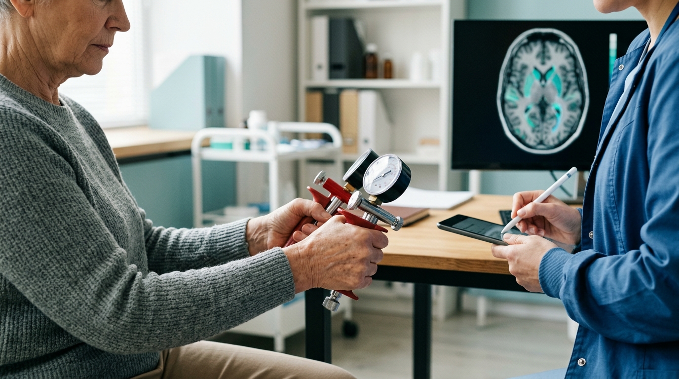
The researchers enrolled 353 older adults from the Magdeburg area of Germany who showed no signs of cognitive impairment at the time of the study. The sample’s mean age was 72.77 years, with a standard deviation of 7.95 years; 177 of the 353 participants were women. All participants had normal or corrected-to-normal vision and no current or recent psychiatric or neurological diagnoses. The study was conducted at the German Center for Neurodegenerative Diseases (DZNE) in Magdeburg and Martin Luther University Halle-Wittenberg. (Schwarck et al., npj Aging, 2026)
The researchers measured two distinct categories of physical fitness. Muscular capacity was assessed through three separate measures: handgrip strength, appendicular skeletal muscle mass (the combined mass of the muscles in the arms and legs), and a Timed-Up-and-Go test, which measures how quickly a person can rise from a chair, walk three meters, turn around, walk back, and sit down again. It is a standard functional measure in geriatric research because it captures not just strength but coordination, balance, and the integrated efficiency of the motor system.
Aerobic fitness was measured using VO2max — the maximum rate at which the body can consume oxygen during exercise. Because exercise testing involves cardiovascular stress, only participants who met specific safety criteria were tested for VO2max; 140 of the 353 participants in the full sample took part in this portion of the assessment. This means the VO2max group was a healthier subset — a limitation the researchers acknowledge directly.
Brain imaging included measures of gray matter volume and cortical thickness, assessment of perivascular spaces in the basal ganglia, and tau-PET imaging to detect early tau protein accumulation in the medial temporal lobe — a region closely associated with episodic memory. White matter hyperintensities were also measured. Blood-based biomarkers for neurodegeneration were assessed in a smaller subsample.
What Aerobic Fitness Showed in the Brain
Among the 140 participants tested for aerobic capacity, higher VO2max scores were associated with greater medial temporal lobe cortex thickness and greater total gray matter volume. The medial temporal lobe — which includes the hippocampus and surrounding cortex — is a region involved in the formation and retrieval of episodic memories and is among the first areas to show structural changes in neurodegenerative conditions. Greater thickness in this region, in a sample of people who show no cognitive impairment, suggests that aerobic fitness may be associated with preserved structural integrity of tissue that would otherwise decline. (Schwarck et al., npj Aging, 2026)
Total gray matter volume reflects the overall quantity of neuron-containing tissue across the brain. In older adults, gray matter volume tends to decrease with age; associations between aerobic fitness and preserved gray matter volume have appeared in earlier research as well, though the mechanism is not fully established. The researchers in this study did not find an association between VO2max and tau accumulation in the medial temporal lobe, suggesting that aerobic fitness may support structural maintenance without directly slowing protein pathology in this region.
The researchers also examined whether aerobic fitness was associated with cognitive performance on a battery of tests. They found that VO2max added only 7.6 percent of additional variance in delayed verbal recall beyond what was already explained by tau burden in the medial temporal lobe. This is a small contribution. The structural associations were present; the direct cognitive benefit in this sample was modest. The researchers suggest this is consistent with a brain maintenance model rather than a cognitive reserve model — meaning fitness may help preserve tissue, but the research does not show it compensating for or outpacing existing pathology.
What Muscular Capacity Showed — and Why the Location Surprised Researchers
The muscular capacity findings pointed to a different region of the brain entirely. Better performance across the three muscular measures — handgrip strength, appendicular skeletal muscle mass, and Timed-Up-and-Go — was associated with lower perivascular space volume in the basal ganglia. The basal ganglia are a cluster of structures deep in the brain involved in motor coordination, habit formation, and the regulation of movement. They were not the structures researchers typically look to first when studying fitness and memory.
Perivascular spaces — the subject of the next section — are not visible on the kinds of brain imaging many people have heard of. They require specific MRI protocols to detect and quantify, and their relationship to brain health is an active area of research. Finding that muscular capacity connects to this specific measure, in this specific region, was one of the more unexpected findings of the study. (Schwarck et al., npj Aging, 2026)
It is important to be precise about what “lower perivascular space volume” means here and what it does not mean. In the context of this study, smaller perivascular spaces in healthy older adults who show no cognitive impairment are associated with better muscular capacity. The researchers interpret this as a potential indicator of more efficient brain waste clearance in those individuals. This interpretation is plausible and grounded in current understanding of what perivascular spaces do — but it is an inference, not a directly measured finding in this study. The researchers note that muscular capacity findings should be interpreted with caution, because measures of skeletal muscle mass and grip strength can be influenced by body composition in ways that are difficult to fully control for.
A separate meta-analysis published in 2021 in Frontiers in Aging Neuroscience (Cui et al., PMC7890203) examined 15 longitudinal cohort studies comprising 27,588 individuals and found that lower grip strength was associated with a hazard ratio of 1.99 for cognitive decline, 1.54 for any dementia, and 1.41 specifically for Alzheimer’s disease. Those are associations over time in observational data — they do not establish what mechanism links muscle strength to cognitive outcomes. The 2026 study offers one candidate mechanism: the perivascular space pathway.
What Perivascular Spaces Do — and Why Their Size Matters
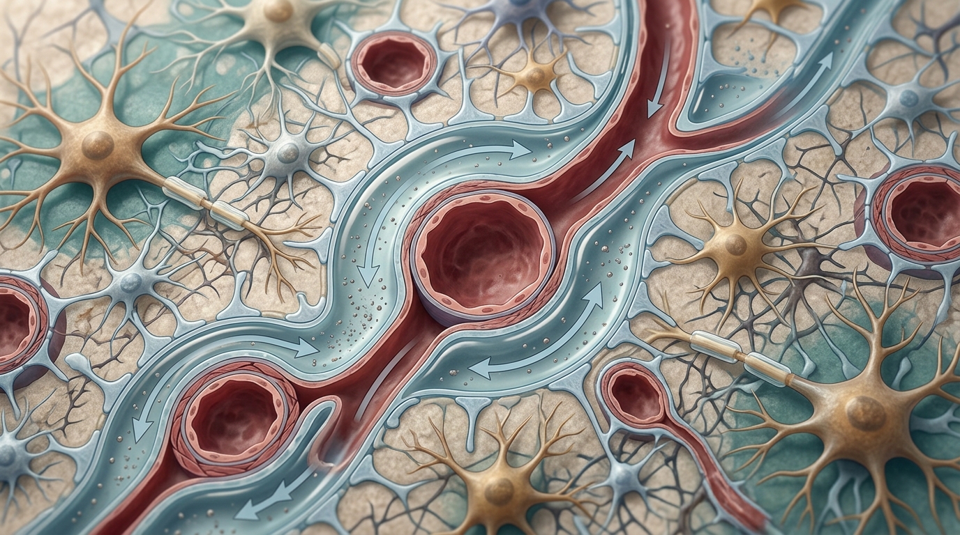
Perivascular spaces are microscopic fluid-filled channels that surround the blood vessels running through brain tissue. They are part of what researchers call the glymphatic system — a waste-clearance network that uses the pulsation of blood vessels and the flow of cerebrospinal fluid to flush metabolic byproducts out of the brain. The name comes from glial cells, particularly astrocytes, whose end-feet line the outer walls of these spaces and regulate fluid movement through them.
Among the substances this system helps clear are soluble forms of amyloid-beta and tau — the proteins whose accumulation in the brain is associated with Alzheimer’s disease and related conditions. A 2021 review in Neuroradiology by Gouveia-Freitas and Bastos-Leite examined the role of perivascular spaces in brain waste clearance and found that enlarged perivascular spaces correlate with impaired beta-amyloid removal. The spaces tend to enlarge with age; when they become abnormally enlarged, the enlargement is considered a marker of underlying dysfunction in the waste-clearance system rather than a feature of efficient clearance. (Gouveia-Freitas & Bastos-Leite, Neuroradiology, 2021)
The glymphatic system does most of its work during sleep, when brain cells shrink slightly and the spaces between them expand, allowing cerebrospinal fluid to flow more freely. This is one of the reasons sleep deprivation has been associated with increased amyloid-beta accumulation in the brain — the nightly clearing process is interrupted or reduced. In the context of the 2026 fitness study, the finding that muscular capacity associates with smaller perivascular spaces suggests — cautiously — that physically stronger older adults may have a waste-clearance system that is functioning with greater efficiency. What drives that association biologically, and whether intervening to improve muscular capacity in older adults changes perivascular space volume over time, are questions the cross-sectional study design cannot answer.
The Cognitive Performance Finding — and What It Means
One of the most important findings in the 2026 study was what was not found. Despite the clear associations between fitness and brain structure — VO2max with gray matter volume and MTL thickness, muscular capacity with basal ganglia PVS volume — neither fitness type was directly associated with scores on cognitive tests, including global cognition and episodic memory. The VO2max association with delayed verbal recall explained only 7.6 percent of additional variance beyond tau burden alone.
The researchers frame this result carefully. They describe it as consistent with a brain maintenance hypothesis rather than a cognitive reserve hypothesis. Brain maintenance, in this framework, means that fitness is associated with preserving the structural integrity of brain tissue — keeping gray matter volume from declining as steeply, keeping waste-clearance channels functioning more efficiently. Cognitive reserve, by contrast, refers to a hypothesis in which accumulated resources — education, cognitively demanding work, physical fitness — allow a person’s cognition to remain relatively functional even as underlying brain pathology accumulates. The two hypotheses are not mutually exclusive, but the current study’s data fit the maintenance picture better than the reserve picture: the fitness associations showed up in structure, not in cognitive scores.
This distinction matters for how the findings should be interpreted. Brain maintenance is a process that, if supported over decades, may help delay the emergence of cognitive changes — not by compensating for damage after it occurs, but by preserving the tissue that cognitive function depends on. That is a meaningful finding. It is also a different finding from showing that physically stronger or fitter older adults remember more, solve problems faster, or are protected against dementia. The study’s data do not support those stronger claims.
What This Research Does Not Establish
The 2026 study is careful about its own limitations, and those limits are worth stating plainly.
The study design is cross-sectional — it took measurements at one point in time. It can describe associations between fitness and brain structure, but it cannot determine which came first. It is possible that people with healthier brains engage in more physical activity; it is possible that physical activity produces the brain differences; it is possible that both arise from a third factor such as cardiovascular health or genetics. A longitudinal intervention study — measuring the same people before and after a fitness program — would be needed to test whether improving fitness changes brain structure. That study does not yet exist in the form that would answer this question definitively.
The sample was highly educated, predominantly Caucasian, and recruited from one region of Germany. Research consistently finds that education, ethnicity, baseline health, and geography interact with brain aging in complex ways. Results from a highly educated older German cohort may not generalize to populations with different educational histories, different baseline health profiles, or different cultural contexts around exercise.
The VO2max subsample of 140 participants met additional exercise-safety criteria that the full sample did not — meaning the aerobic fitness group was likely healthier on average than the muscular capacity group. Comparing findings across these two groups requires caution because the populations being compared are not the same.
Some of the brain imaging measures, including the tau-PET and blood biomarkers, were collected only in smaller subsamples within the 353. The findings from those smaller groups carry wider uncertainty than the findings from the full sample.
Finally, all of the results describe group-level averages. The hazard ratios in the grip strength meta-analysis and the associations in the brain imaging study describe patterns across thousands of participants. They say nothing about what will happen in any individual. Individual variation in genetics, activity history, health status, sleep, and dozens of other factors means the range of outcomes within any fitness category is wide. Group statistics do not predict individual trajectories.
Frequently Asked Questions
Does physical exercise actually protect the aging brain?
Research consistently finds associations between physical fitness and measures of brain structure in older adults. A 2026 study of 353 cognitively unimpaired older adults found that higher aerobic fitness was associated with greater gray matter volume and thicker medial temporal lobe cortex, while better muscular capacity was associated with smaller perivascular spaces in the basal ganglia. These are associations in a cross-sectional study — they describe a relationship, not a proven causal direction. The study found that fitness was not directly associated with cognitive performance scores, leading the researchers to describe their findings as consistent with brain maintenance rather than cognitive reserve.
What is a perivascular space, and why does its size matter in brain aging?
Perivascular spaces are fluid-filled channels that surround the blood vessels running through brain tissue. They are part of the brain’s glymphatic waste-clearance system, which removes metabolic byproducts — including soluble forms of amyloid-beta and tau — from brain tissue, primarily during sleep. In older adults, these spaces tend to enlarge, and pathological enlargement is considered a marker of reduced efficiency in this waste-clearance system. The 2026 Schwarck et al. study found that better muscular capacity (grip strength, skeletal muscle mass, Timed-Up-and-Go performance) was associated with smaller perivascular spaces in the basal ganglia, suggesting a possible connection between muscular fitness and the efficiency of the brain’s waste-clearance pathway.
Is aerobic exercise or strength training more important for brain health?
The 2026 study found that aerobic fitness and muscular capacity were each associated with different aspects of brain structure in older adults, through distinct pathways. Aerobic fitness (VO2max) showed associations with gray matter volume and medial temporal lobe thickness. Muscular capacity showed associations with perivascular space volume in the basal ganglia. The two types of fitness were not compared head-to-head in terms of which is more important. What the study suggests is that they may contribute to brain health through different mechanisms, rather than one being superior. This is a cross-sectional association study; it cannot prescribe which type of exercise prevents cognitive decline in individuals.
If fitness was associated with better brain structure, why wasn't it associated with better memory scores?
This was one of the unexpected findings of the 2026 study. The researchers found structural associations — fitness connected to gray matter and perivascular spaces — but not direct cognitive performance associations. They interpret this as consistent with a brain maintenance hypothesis: fitness may help preserve brain tissue integrity over time, rather than compensating for existing pathology or directly boosting cognitive performance in a cross-sectional snapshot. In this sample of cognitively unimpaired older adults, the variation in cognitive scores may have been too small to detect a fitness effect. Longer-term longitudinal studies would be better suited to testing whether fitness-related brain maintenance translates into delayed cognitive decline over years.
Do these findings apply to all older adults?
The 2026 study enrolled highly educated, predominantly Caucasian older adults from one region of Germany, and the VO2max subsample met additional exercise-safety criteria that made them a healthier subgroup. The findings describe associations within this specific population and may not generalize directly to older adults with different educational backgrounds, health histories, or physical baselines. The grip strength meta-analysis (Cui et al., 2021) drew on 27,588 individuals across 15 longitudinal studies from varied populations, which increases confidence in the general association between muscular fitness and cognitive outcomes — but it also works with group-level hazard ratios that describe population patterns, not individual predictions. Both lines of evidence are informative; neither dictates what will happen to any particular person.
Sources
- Schwarck S et al. Associations of physical fitness with brain structure, pathology and cognition in cognitively normal older adults (2026) — npj Aging (Nature Portfolio)
- Cui M et al. Grip Strength and the Risk of Cognitive Decline and Dementia: A Systematic Review and Meta-Analysis of Longitudinal Cohort Studies (2021) — Frontiers in Aging Neuroscience / PubMed Central
- Gouveia-Freitas K, Bastos-Leite AJ. Perivascular spaces and brain waste clearance systems: relevance for neurodegenerative and cerebrovascular pathology (2021) — Neuroradiology / PubMed Central
Understand Your Nervous System
The relationship between physical activity, brain structure, and cognitive function across the lifespan is one of the areas we follow closely at NeuroBalance. If you are curious about how brain regulation and nervous system function connect to your own experience of aging, attention, or stress, we offer a free initial consultation. Our practice is private, one-to-one, and focused on understanding your specific nervous system — not a protocol applied to a category.
Schedule Your Free Consultation
Call us: (424) 625-5445 · Los Angeles, CA
Disclaimer: This content is for informational purposes only and does not constitute medical advice, diagnosis, or treatment. LENS Neurofeedback is not FDA-approved for all conditions mentioned. Please consult with a qualified healthcare provider before beginning any new treatment program.

A balanced brain is a better brain for a happier life