
Fibromyalgia: What New Research Is Finding Inside the Central Nervous System
Fibromyalgia has been dismissed as 'all in your head' for decades. A 2026 systematic review of neuroimaging and cerebrospinal fluid studies now shows the central nervous system is genuinely involved — though not in any way researchers have fully mapped yet.
For decades, one phrase followed fibromyalgia through clinical encounters with a persistence that felt deliberate: “it’s all in your head.”
It was used as a way to close a conversation rather than open one — to move a patient toward psychiatry when no bloodwork explained the pain, to code a condition as medically unexplained when no structural damage showed on a scan. The phrase carried a clear implication: that fibromyalgia was about imagination, personality, or emotional excess rather than biology.
Researchers studying the central nervous system have spent the last two decades building a case that the phrase was accurate in a way none of its users intended. Fibromyalgia does appear to involve the brain. What neuroimaging, cerebrospinal fluid analysis, and a 2026 systematic review are now beginning to map is what that involvement actually looks like — and what it still does not explain.
What a 2026 Systematic Review Assembled — and What It Found
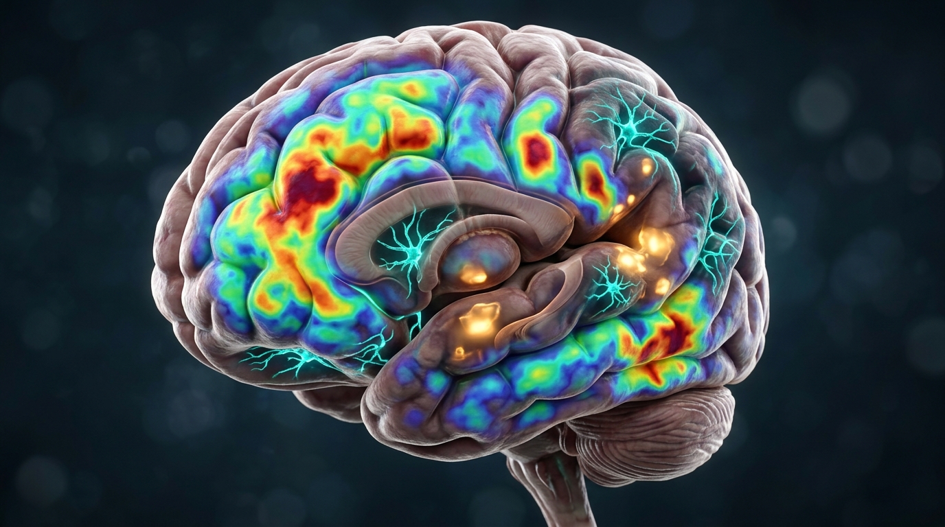
In 2026, researchers Mireia Castán-Sogas and Antonia Fusté-Escolano published a systematic review specifically examining whether there is measurable evidence of central neuroimmune alterations in fibromyalgia. The review, published in Brain, Behavior, and Immunity, searched three major databases — PubMed, Web of Science, and Scopus — for case-control studies comparing fibromyalgia patients to healthy controls on neuroimaging or biological markers of central immune activity.
Eighteen studies qualified for inclusion. Nine used neuroimaging techniques — predominantly positron emission tomography and functional MRI — to look directly at brain activity and structure. Six examined cerebrospinal fluid, the liquid that bathes the brain and spinal cord and which carries markers of what is happening inside the central nervous system. Two analyzed differential gene expression. One used a translational cellular approach.
Despite significant heterogeneity across those studies — they varied in methodology, sample size, imaging tracers, and what they chose to measure — Castán-Sogas and Fusté-Escolano found what they described as convergent patterns. Across different research groups using different methods, similar directions of change were appearing: signs of glial and neurometabolic abnormalities in cortical and subcortical brain regions, alongside alterations in immune-related proteins and markers of disrupted neuroimmune signaling.
The review was conducted in accordance with PRISMA 2020 guidelines and registered in PROSPERO before data collection began — standard practice for systematic reviews intended to be both replicable and transparent about what was searched and what was excluded. The researchers were explicit that the heterogeneity across studies made firm conclusions difficult, and the review is best understood as a map of what has been measured so far, not a settled account of mechanism.
What PET Imaging Has Shown About Glial Cells in the Brain
One of the most striking contributions to the 2026 systematic review’s neuroimaging category came from a study published seven years earlier — and was, at the time, described by its authors as the first multi-site direct evidence of brain glial activation in fibromyalgia.
In 2019, researchers led by Marco Loggia at Massachusetts General Hospital and drawing on a parallel dataset from the Karolinska Institutet in Sweden conducted positron emission tomography scans of 31 fibromyalgia patients and 27 healthy controls. The imaging tracer they used — [11C]PBR28 — binds to a protein called the translocator protein (TSPO), which is upregulated in activated microglia and astrocytes. Microglia are the brain’s resident immune cells. Astrocytes provide structural and metabolic support to neurons. Both become activated under conditions of injury, infection, or sustained neurological stress.
Compared to healthy controls, fibromyalgia patients in the Loggia et al. (2019, Brain, Behavior, and Immunity) study showed widespread cortical elevations in the TSPO marker, most pronounced in the medial and lateral walls of the frontal and parietal lobes. A second tracer targeting astrocytes specifically did not show the same pattern, suggesting the signal was more likely microglial in origin — though the authors noted that larger studies would be needed to fully assess astrocytic contributions.
TSPO binding is not a perfect proxy for neuroinflammation. The protein is expressed in several cell types and under various conditions, and elevated TSPO does not definitively confirm a specific inflammatory pathway. But the pattern — widespread, cortical, and not observed in healthy controls — added a new category of measurement to the fibromyalgia research landscape: one that could actually look at what was happening inside the brain rather than inferring it from pain reports or behavioral tests.
What Cerebrospinal Fluid Analysis Has Documented
While imaging techniques offer a window into brain structure and activity, cerebrospinal fluid analysis offers something different: a chemical readout of what is circulating in direct contact with the brain and spinal cord.
Among the most consistent findings in fibromyalgia research is an elevation of substance P in the cerebrospinal fluid of fibromyalgia patients. Substance P is a neuropeptide that functions as a pain-signaling molecule — it is released in response to noxious stimuli and helps transmit pain signals through the spinal cord. Measurements of cerebrospinal fluid in fibromyalgia patients have found approximately a threefold elevation in substance P compared to healthy controls, alongside a roughly fourfold increase in nerve growth factor.
The 2026 systematic review by Castán-Sogas and Fusté-Escolano noted that among the six cerebrospinal fluid studies it included, changes in the concentrations of cytokines and other immune molecules appeared alongside these neuropeptide alterations — markers of immune signaling rather than classical neurotransmission. Alterations in the expression of genes related to inflammatory processes were also documented in the gene expression studies.
Cerebrospinal fluid studies are not collected easily. The analysis requires a lumbar puncture under controlled conditions, which limits the sample sizes available to researchers and makes replication more expensive. Most of the studies in the systematic review had relatively small participant groups, which is one reason the review’s authors emphasized the heterogeneity across their included studies and the difficulty of drawing firm conclusions from any single finding.
What the CSF evidence adds, taken together with the imaging findings, is a picture of the central nervous system environment in fibromyalgia that differs measurably from healthy controls in multiple biological registers — not just in how patients report pain, but in what is circulating in the space where the brain and spinal cord live.
Central Sensitization: When the Nervous System's Gain Is Set Too High
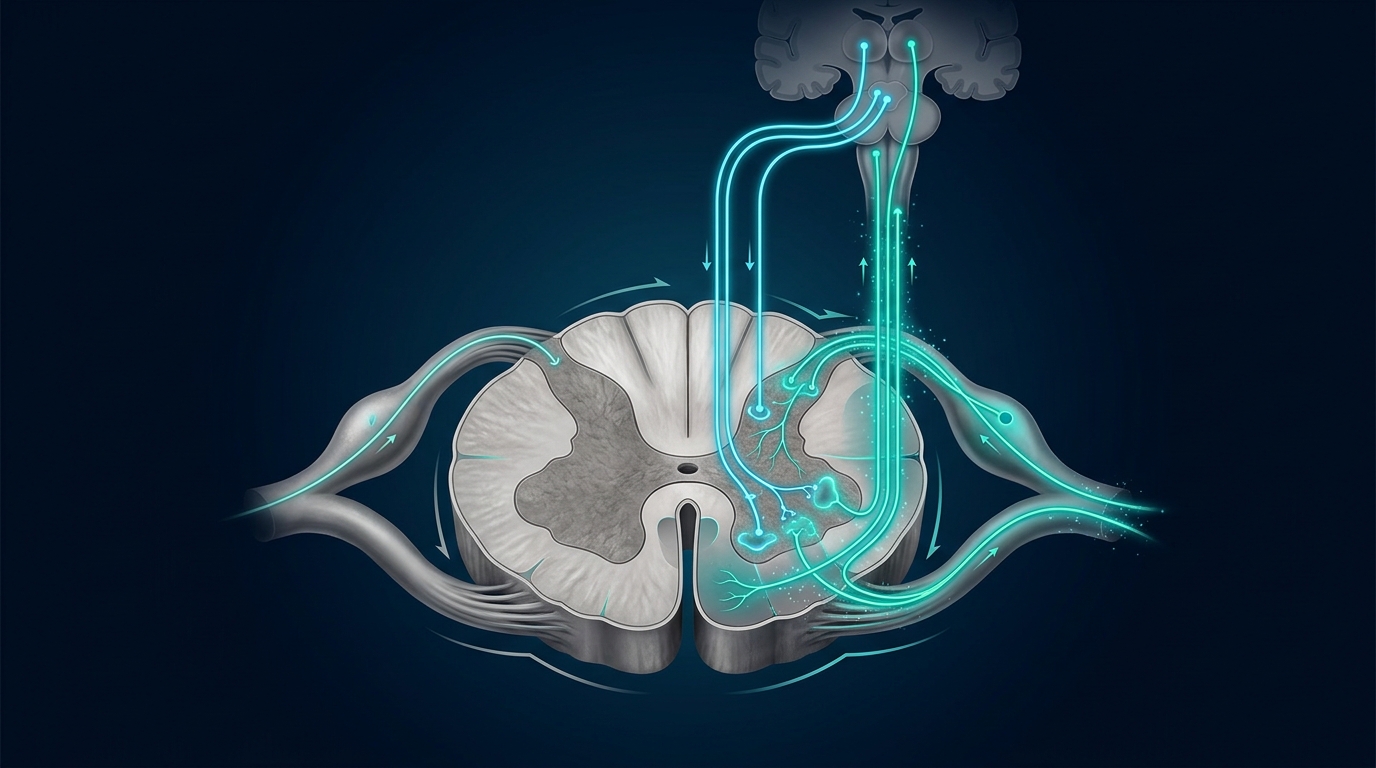
One framework that has become central to understanding fibromyalgia — and that the neuroimmune research above helps to explain biologically — is central sensitization. The concept describes a state in which the central nervous system’s pain-processing circuitry becomes amplified: signals that would not normally register as painful produce pain, and signals that are painful produce a level of pain that seems disproportionate to the tissue state.
Under normal conditions, the nervous system both amplifies pain signals (ascending nociception — the system that warns the brain about damage) and suppresses them (descending inhibition — the system that modulates incoming signals and prevents the brain from being overwhelmed). In central sensitization, ascending nociception is increased and descending inhibition is impaired. The result is something like a volume control set permanently higher than it should be, with the dampening circuit partially disabled.
At the neurochemical level, research has identified elevated glutamate in the central nervous systems of people with fibromyalgia. Glutamate activates NMDA receptors, which are involved in amplifying pain signals over time — a process called temporal summation, where repeated low-level stimuli add up to create a disproportionate pain response. Inhibitory neurotransmitters, including serotonin and met-enkephalin, appear reduced in fibromyalgia cerebrospinal fluid compared to controls.
The glial activation seen in the PET imaging, and the immune molecule changes seen in CSF studies, are consistent with this sensitization picture. Activated microglia release pro-inflammatory cytokines that can lower the threshold at which neurons fire. If the 2026 systematic review’s evidence of glial and neurometabolic abnormalities holds up across larger and more methodologically consistent studies, it would suggest that the sensitization in fibromyalgia may not simply be a matter of misaligned neurotransmitter ratios — it may involve an ongoing immune-like process in the brain itself.
That distinction has implications for how the biology is understood, even if it does not yet change what can be done about it clinically.
A Pattern Across the Evidence — and Its Limits
The picture that emerges from the 2026 systematic review, the earlier PET imaging research, and the cerebrospinal fluid literature is one of consistent directional signals across methodologically diverse studies. Multiple research groups, using different instruments and measuring different markers, have found the central nervous system in fibromyalgia patients to differ from healthy controls in overlapping ways.
Neuroimaging studies have found elevated markers of glial activity in cortical regions involved in pain processing and attention. Cerebrospinal fluid studies have found elevated pain-signaling molecules and immune-related proteins. Gene expression studies have found changes in inflammatory pathways. The 2026 review by Castán-Sogas and Fusté-Escolano described these as “convergent patterns” — not agreement on the exact mechanism, but multiple measurement types pointing in the same direction.
For the many people who have been told their symptoms are unexplained or psychogenic, this body of evidence is meaningful. It represents a measurable, replicable departure from the biology of people without fibromyalgia, visible in the brain’s own tissue and in the fluid that surrounds it — not derived from questionnaires or self-report alone.
That is a different kind of claim than what the research can support if pushed further. The studies included in the systematic review ranged considerably in sample size, in which brain regions they examined, and in whether they found the same markers elevated by the same degree. High heterogeneity across studies is the review’s own characterization. The biology is real and measurable; its specific pathway, magnitude, and clinical significance remain areas of active investigation rather than settled science.
What This Research Does Not Establish — The Honest Accounting
The convergent signals in this literature are real. The limitations are also real, and the researchers most closely involved in this work are generally careful about what they claim.
The most fundamental open question is causal direction. Fibromyalgia involves chronic, widespread pain. Chronic pain of any kind is known to change the central nervous system — it induces neuroplastic changes, alters glial activity, and modifies the biochemical environment of the brain over time. The studies in the 2026 systematic review are case-control designs: they compared people with fibromyalgia to healthy controls at a single point in time. They were not designed to determine whether the CNS changes preceded the pain and contributed to it, or developed in response to years of chronic pain, or both.
This is not a trivial distinction. If central neuroimmune alterations are primarily a downstream consequence of chronic pain rather than a driving cause, the clinical implications are different than if they are upstream contributors.
Second, none of the biological markers identified so far constitutes a clinical diagnostic tool for fibromyalgia. No blood test, no CSF marker, no PET scan pattern reliably identifies fibromyalgia in an individual — these are group-level findings from research populations, and the overlap with healthy controls in many studies is substantial. Fibromyalgia remains a clinical diagnosis based on the presence and distribution of symptoms.
Third, sample sizes across the included studies were often small. The systematic review included 18 studies total across all measurement types — with some individual studies enrolling fewer than 20 participants in each group. Small samples produce noisier estimates, are more vulnerable to chance findings, and make it harder to detect which brain regions and biological pathways are genuinely involved versus artifacts of the specific participants studied.
The value of the 2026 systematic review is that it consolidated what has been measured so far and described the overall pattern honestly, including its heterogeneity. The honest summary of that pattern: there is consistent evidence that the central nervous system in fibromyalgia differs measurably from healthy controls across multiple biological markers — and insufficient evidence to say precisely how, why, or in which causal direction those differences operate.
Frequently Asked Questions
What did the 2026 systematic review find about fibromyalgia and the central nervous system?
The review by Castán-Sogas and Fusté-Escolano (Brain, Behavior, and Immunity, 2026) examined 18 case-control studies comparing fibromyalgia patients to healthy controls on neuroimaging and biological markers of central immune activity. Despite high heterogeneity across methods, the review identified convergent patterns: glial and neurometabolic abnormalities in cortical and subcortical brain regions, changes in immune-related proteins in cerebrospinal fluid, and alterations in genes related to inflammatory processes. The authors characterized the findings as consistent directional signals across diverse methodologies rather than a definitive established mechanism.
What is glial activation and why does it matter for understanding fibromyalgia?
Glial cells — including microglia and astrocytes — are the brain’s non-neuronal support and immune cells. Microglia in particular function as the brain’s resident immune system; when activated, they release pro-inflammatory cytokines that can lower the threshold at which neurons fire and amplify pain signaling. A 2019 multi-site PET imaging study at Massachusetts General Hospital and the Karolinska Institutet found widespread cortical elevations in a glial activation marker in 31 fibromyalgia patients compared to 27 healthy controls — the first multi-site direct imaging evidence of this kind. Whether this activation drives fibromyalgia symptoms, develops in response to chronic pain, or both, is not yet established.
What does central sensitization mean, and how is it connected to fibromyalgia?
Central sensitization describes a state in which the central nervous system’s pain-processing becomes amplified — ascending pain signals are increased while descending inhibitory controls are reduced. Stimuli that would not normally be painful can trigger pain (allodynia), and stimuli that are painful produce a disproportionate response (hyperalgesia). Research in fibromyalgia has found elevated levels of substance P (a pain-signaling neuropeptide, roughly 3-fold elevated in cerebrospinal fluid) and glutamate, alongside reduced inhibitory neurotransmitters. Central sensitization is the current leading framework for understanding why widespread pain occurs without identifiable tissue damage.
Does this research prove that fibromyalgia is caused by neuroinflammation?
No. The research identifies consistent biological differences between fibromyalgia patients and healthy controls in the central nervous system — measurable in imaging, cerebrospinal fluid, and gene expression — but it does not establish the causal direction of those differences. Chronic pain of any kind can produce neuroplastic changes and alter glial activity over time, meaning the CNS differences observed could be a consequence of years of chronic pain rather than its initiating cause. The studies are case-control designs that compare groups at a single point in time and are not designed to determine which came first. None of the biological markers identified constitutes a clinical diagnostic test.
What do these findings mean for someone experiencing symptoms associated with widespread pain?
For people who have encountered skepticism about whether their symptoms are ‘real,’ the cumulative evidence from neuroimaging, cerebrospinal fluid studies, and the 2026 systematic review represents a body of measurable biology — findings that are not derived from self-report alone. The central nervous system in fibromyalgia shows consistent, reproducible differences from healthy controls across multiple research groups and methods. What that biology means for any individual’s experience, trajectory, or response to different approaches remains highly variable and is not something population-level research can predict.
Sources
- Castán-Sogas & Fusté-Escolano (2026) — Evidence of central neuroimmune alterations in fibromyalgia: a systematic review of case-control studies — Brain, Behavior, and Immunity, 136, 106773
- Loggia et al. (2019) — Brain glial activation in fibromyalgia: A multi-site positron emission tomography investigation — Brain, Behavior, and Immunity, 75, 72–83
- Neuroscience News — Widespread Inflammation in Brains of Those with Fibromyalgia — Neuroscience News
- News-Medical — Study links neuroimmune dysregulation to complex fibromyalgia symptoms (2026) — News-Medical
- Neuroinflammatory and Immunological Aspects of Fibromyalgia (2025 review) — PMC / National Library of Medicine
Understand Your Nervous System
Brain biology is complex, and the research into conditions like fibromyalgia is still mapping territory that was considered unmeasurable a generation ago. If you're trying to understand what's happening in your own nervous system, one-to-one sessions with a trained practitioner can offer something population research cannot: attention to the specific patterns of your brain, not averages across a study group.
Schedule Your Free Consultation
Call us: (424) 625-5445 · Los Angeles, CA
Disclaimer: This content is for informational purposes only and does not constitute medical advice, diagnosis, or treatment. LENS Neurofeedback is not FDA-approved for all conditions mentioned. Please consult with a qualified healthcare provider before beginning any new treatment program.

A balanced brain is a better brain for a happier life.