
Is Your Brain Older Than You Are? The New Science of 'Brain Age'
Researchers can now estimate a brain’s age from structural MRI data — and it often differs from chronological age. A 2025 study of more than 41,000 participants found the gap predicts Alzheimer’s risk, mortality, and a range of lifestyle factors. Here is what the evidence shows, and where it honestly stops.
Most people carry a quiet assumption about their brain: that it ages at roughly the same pace as the rest of them. If you are 52, your brain is 52. The idea that chronological age and brain age could diverge significantly — that a 50-year-old might have a brain whose structural properties resemble those of someone ten years older, or younger — is not a framework most people apply to themselves.
Neuroscience has been quietly building that framework for about fifteen years. The approach is called brain age estimation, and it works by training machine-learning models on brain scan data from thousands of people across a wide age range, then using those models to predict the age of a new brain from its structural properties. The difference between the model’s prediction and a person’s actual age is called the brain age gap, or BAG — and it has emerged as one of the more actively researched biomarkers in the study of aging, mental health, and neurodegenerative disease.
This article examines what that gap is, what the current evidence associates it with, and — equally important — what it does not yet establish.
What 'Brain Age' Actually Measures
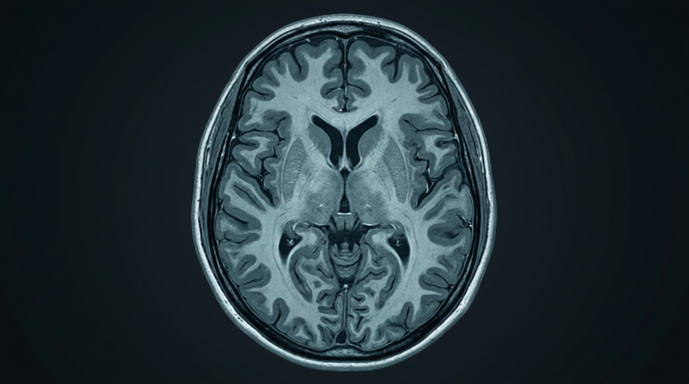
A structural MRI produces a detailed image of the brain — its gray matter volume, white matter density, cortical thickness, and the relative sizes of subcortical structures. None of these features is directly labeled young or old, but they follow recognizable patterns across the lifespan. Gray matter volume tends to decline gradually with age. White matter shows characteristic changes in density and integrity. The thickness of the cortical surface changes across regions at different rates.
Machine-learning models trained on large samples of healthy brains can learn to predict chronological age from these features with measurable accuracy. A 2024 comprehensive review in Tomography (Wu et al.) examined 52 machine-learning and deep-learning studies on brain age estimation published between 2020 and 2024. It found that T1-weighted structural MRI was the dominant imaging modality and that models using whole-brain structural features consistently outperformed those relying on individual regions alone (Wu et al., Tomography, 2024).
The accuracy of these models is typically reported as mean absolute error — the average difference in years between predicted and actual age. In the largest studies, this figure has come down to around 2 to 3 years. A 2025 study published in Communications Medicine by Zhang and colleagues trained a 3D Vision Transformer model on data from 41,551 participants across 13 UK Biobank datasets and achieved a mean absolute error of 2.68 years (Zhang et al., Communications Medicine, 2025). Predicting within less than three years on average, at this scale, is what makes the brain age gap a potentially informative measurement rather than a rough approximation.
The gap itself — predicted age minus chronological age — is the quantity researchers examine. A positive brain age gap means the brain’s structural properties resemble those of someone older. A negative gap means they resemble someone younger. A gap of zero means the model’s estimate and the calendar agree.
What the Brain Age Gap Predicts
The Zhang et al. 2025 study is currently the largest systematic examination of what the brain age gap predicts. Across 41,551 participants, the researchers found that a one-year increase in the brain age gap was associated with a 16.5% increase in Alzheimer’s disease risk and a 12% increase in all-cause mortality risk. Participants in the highest quartile for brain age gap had 2.8 times the Alzheimer’s disease risk of those in the lowest quartile. The model also found associations with a range of lifestyle factors — sleep quality, physical activity, alcohol use, and BMI — and with psychiatric conditions including depression, anxiety, and PTSD (Zhang et al., Communications Medicine, 2025).
These are significant associations. They place the brain age gap in a statistical relationship with outcomes that matter — dementia risk, mortality, mental health — and suggest that the gap is capturing something real about biological aging rather than noise.
The lifestyle associations are particularly worth noting. Higher body mass index, poorer sleep quality, greater alcohol consumption, and lower physical activity were each associated with a larger brain age gap — a brain whose structure appeared older relative to calendar age. The associations ran in both directions: factors generally considered protective for health were associated with a younger-appearing brain. This bidirectional consistency across multiple lifestyle variables in a single large dataset adds to the picture of the brain age gap as a meaningful summary measure of biological aging.
The association with psychiatric conditions — depression, anxiety, PTSD — raises a question the study does not resolve: does psychological distress age the brain, does a biologically older brain create vulnerability to those conditions, or do both reflect shared upstream factors? The association establishes that they travel together. It does not establish which way the influence runs.
Depression, Aging, and the Brain Age Gap
The relationship between depression and brain aging has received specific attention. A 2026 study in Psychological Medicine (Mitchell et al.) examined brain age prediction in a sample of 190 participants, comparing those with current or lifetime depression diagnoses against healthy controls across a range of ages.
The primary finding was an age-by-group interaction (beta = 0.34, p < 0.001): older adults with depression showed a significantly greater brain age gap than older controls, while the difference was not significant in younger adults. There was no main effect of depression on brain age across the whole sample — the association between depression and an older-appearing brain was specific to older individuals in this cohort.
The study also examined cortisol as a potential mediating variable. Elevated cortisol is often proposed as a biological pathway by which chronic stress and depression might accelerate brain aging — a reasonable hypothesis given that prolonged HPA axis activation is associated with structural changes in brain regions including the hippocampus. The Mitchell et al. findings, however, showed that cortisol was negatively correlated with the brain age gap in this sample: higher cortisol was associated with a smaller gap, the opposite of what was predicted. The researchers noted that acute cortisol sampling may not capture the chronic HPA activation that is most relevant to cumulative structural change, and that the relationship may be more complex than a simple linear association (Mitchell et al., Psychological Medicine, 2026).
The cortisol finding is worth sitting with precisely because it did not come out as expected. It is a reminder that even within this research area, the biological mechanisms that connect psychological and structural aging are not fully understood. The association between depression and an older-appearing brain in older adults is real in this sample, but the pathway remains unclear.
What the Brain Age Gap Does Not Tell You
This is where careful reading of the evidence matters as much as the findings themselves.
The brain age gap is a research construct, not a clinical diagnostic tool. No standard clinical neuroimaging workflow produces a brain age estimate for individual patients. There is no validated reference range that would allow a clinician to determine whether a particular person’s brain age gap is medically concerning or how to act on it. The Wu et al. 2024 review noted that training datasets are predominantly drawn from European populations, skewed toward healthy adults, with relative underrepresentation of diverse demographic groups — a limitation that affects how well models generalize beyond their training samples (Wu et al., Tomography, 2024).
The Zhang et al. 2025 study is observational. It documents associations between brain age gap and health outcomes in a very large sample, but it cannot establish that a larger brain age gap causes disease. The causal arrow could run in either direction: early-stage disease processes, including early Alzheimer’s pathology, could change brain structure in ways that make the brain appear older to the model. Or both the brain age gap and the health outcomes could reflect a third variable that influences both. Longitudinal studies that follow individuals over time are better positioned to address directionality, and they are ongoing.
The Mitchell et al. 2026 study had 190 participants — carefully designed, but modest in scale relative to the complexity of the interactions being investigated. The cortisol finding, which ran contrary to prediction, underscores how much remains unresolved about the mechanism connecting mood, stress hormones, and structural brain change.
There is also a fundamental limit to what a single structural scan captures. The brain age gap is derived from how a brain looks on one MRI at one point in time. It does not measure the dynamics of how brain circuits communicate, the accumulation of specific proteins, metabolic activity, or the history of inflammatory activity in the brain’s immune cells. Two people with the same brain age gap could have arrived there by different routes and face different biological risks going forward. The gap summarizes; it does not diagnose.
What the Research Establishes, and Where It Is Honestly Incomplete
The brain age research field has produced findings substantive enough to warrant serious attention. A study of 41,551 participants showing a 16.5% increase in Alzheimer’s disease risk per year of brain age gap is not a marginal result. The consistent associations with modifiable lifestyle factors — sleep, physical activity, alcohol use, body weight — align with what is known from other lines of research about what contributes to healthy brain aging. The depression-brain-age interaction in older adults, while drawn from a smaller sample, raises questions that other research groups are actively investigating.
What it does not yet establish is a clinical application. No consensus exists on which brain age model to use clinically, what population it should be validated against, what constitutes a meaningful gap, or what intervention — if any — would be appropriate in response to a particular result. Researchers are working toward these questions; the field has not yet answered them.
The statistic of 16.5% increased risk per year of brain age gap describes a population-level relationship across more than 41,000 people. It does not describe the trajectory of any individual brain, or what a particular person’s brain age gap would mean for their own risk. Group-level statistics characterize distributions; they do not translate directly into individual predictions. That is not a limitation unique to brain age research — it applies to most population-derived biomarkers — but it is a limit worth stating clearly rather than glossing over.
The honest picture that emerges from this research is something like this: the structural properties of the brain, as captured on an MRI, carry more information about biological aging than the number of years on a calendar. That information is real, measurable, and connected to outcomes that matter. It is also being examined at a level of resolution — population studies, observational designs, single imaging modalities — that supports confident associations but not confident individual clinical guidance. The gap between what the science shows and what it would take to act on it in a clinical setting is the work now in progress.
Frequently Asked Questions
What is the brain age gap?
The brain age gap is the difference between a machine-learning model’s estimate of a brain’s structural age — based on features like gray matter volume, cortical thickness, and white matter density from an MRI scan — and a person’s actual chronological age. A positive gap means the brain’s structural properties resemble those of someone older; a negative gap means they resemble someone younger.
What does a larger brain age gap predict?
In a 2025 study of 41,551 participants (Zhang et al., Communications Medicine), each one-year increase in brain age gap was associated with a 16.5% increase in Alzheimer’s disease risk and a 12% increase in all-cause mortality. Participants with the largest brain age gaps had 2.8 times the Alzheimer’s risk of those with the smallest. The gap was also associated with modifiable factors including sleep quality, physical activity, alcohol use, and BMI. These are population-level associations, not individual predictions.
Does depression affect brain age?
A 2026 study in Psychological Medicine (Mitchell et al.) found that older adults with depression had a significantly larger brain age gap than older healthy controls, but this difference was not statistically significant in younger adults. The study did not establish a causal pathway, and a cortisol finding ran contrary to the researchers’ predictions, leaving the biological mechanism unclear. The relationship between mood disorders and brain aging is an active research area without a final answer.
Can lifestyle choices affect the brain age gap?
The Zhang et al. 2025 study found associations between the brain age gap and several lifestyle factors — worse sleep quality, less physical activity, higher alcohol consumption, and higher BMI were each associated with a larger gap. These associations are consistent with other lines of research on brain health, but the study design cannot establish that changing a lifestyle factor will reduce the gap. Randomized trials would be needed to make that claim.
Can I have my brain age measured at a clinic?
Brain age estimation is currently a research tool, not a standard clinical measurement. No clinical workflow produces a validated brain age estimate for individual patients, and no consensus exists on what constitutes a clinically meaningful brain age gap or how a result should guide medical decisions. The research is moving toward clinical application, but that translation has not yet happened.
Sources
- Zhang Y et al. (2025). Brain age gap as a predictive biomarker that links aging, lifestyle, and neuropsychiatric health. Communications Medicine (Lond), 5(1), 188. — Communications Medicine (Nature Portfolio / PMC)
- Mitchell M et al. (2026). Depression and aging: insights from brain age prediction models. Psychological Medicine, 56(1), e103. — Psychological Medicine (Cambridge / PMC)
- Wu Y et al. (2024). Machine Learning and Deep Learning Approaches in Lifespan Brain Age Prediction: A Comprehensive Review. Tomography, 10(8), 1238-1264. — Tomography (MDPI / PMC)
Understand Your Nervous System
If you have been experiencing symptoms associated with cognitive changes, persistent low mood, or difficulty with concentration and memory, a brain health assessment can offer a clearer picture of where your nervous system is currently functioning and identify areas that may benefit from support.
Schedule Your Free Consultation
Call us: (424) 625-5445 · Los Angeles, CA
Disclaimer: This content is for informational purposes only and does not constitute medical advice, diagnosis, or treatment. LENS Neurofeedback is not FDA-approved for all conditions mentioned. Please consult with a qualified healthcare provider before beginning any new treatment program.

A balanced brain is a better brain for a happier life