
What If 'Brain Fog' Isn't One Thing at All?
People recovering from COVID, going through perimenopause, managing poor sleep, and recovering from concussion all describe remarkably similar cognitive symptoms. The experience converges. The biology doesn't.
The people who describe it use remarkably similar language. Thoughts feel thick. Words that should come readily take a moment longer than they should. Reading the same sentence twice and still not fully catching the meaning. A general sense of operating through gauze.
They may have had COVID six months ago. Or they haven’t slept properly in weeks. Or they are in the transition to menopause. Or they took a hard hit to the head playing recreational soccer.
The experience overlaps substantially across these very different situations, and it has acquired an informal name: brain fog. But brain fog is not a clinical diagnosis. It does not appear in any diagnostic manual as a discrete entity. What it is, precisely, is a report — a person describing their experience of cognitive slowing in terms they find accurate.
What changes, depending on who is reporting it and what produced it, is the biology underneath. Following each biological pathway makes the convergence on the same symptom stranger — and more informative — than the shared language suggests.
A Symptom Without a Fixed Address

Brain fog as a complaint has been reported across populations that have almost nothing else in common: people recovering from viral illness, women in the perimenopause transition, individuals with chronic sleep disruption, patients months after a head injury, and people managing autoimmune conditions. Across these groups, the subjective descriptions overlap in ways that go beyond coincidence. The same difficulty accessing words. The same sense of information taking longer to arrive. The same experience of being present in a conversation but not quite in the thought.
But these conditions are not the same biological event. A virus affecting the immune system and a hormonal transition affecting neuroimmune balance are different processes with different time courses and different governing mechanisms. The fact that they produce similar symptoms is a puzzle worth tracing — not because it resolves anything about how to address brain fog, but because understanding where the convergence comes from illuminates something about how cognitive systems respond when their operating conditions are disrupted.
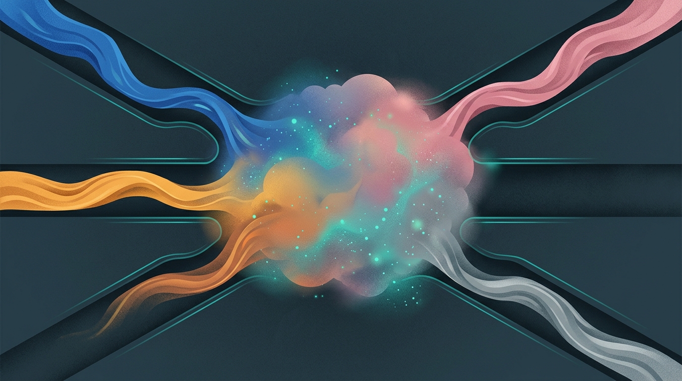
This article follows four pathways from cause to symptom: post-viral immune activation, sleep disruption, hormonal transition, and head injury. Each involves a different starting point, different intermediate steps, and a different biological signature. Each arrives at roughly the same place.
When the Cause Is Viral: The Immune Explanation
A 2026 meta-analysis by Faseeh and colleagues, drawing on 47 studies involving more than 25,000 individuals recovering from COVID-19, found that approximately 30 percent of participants reported brain fog or cognitive impairment. That figure comes from pooled data across studies that measured cognitive complaints at varying time points after infection, using varying definitions of what counts as impairment — a limitation the authors note directly.
What distinguishes post-viral cognitive complaints from other forms of cognitive slowing begins in the immune system. During and after a viral illness, activated immune cells release inflammatory signaling molecules — cytokines — that can cross or influence the blood-brain barrier. In the brain itself, microglia (the brain’s resident immune cells) can shift into reactive states, altering how neural circuits process and transmit information. This immune-mediated disruption is not the same as structural damage: the circuits remain anatomically intact. What changes is the functional environment in which they operate.
The mechanism is: viral trigger → systemic immune activation → cytokine signaling into the central nervous system → microglial reactivity → altered circuit efficiency. The subjective result is a slowing of cognitive processes that feels, from the inside, like thickness or delay.
One important feature of this pathway is that it can persist well past the resolution of the acute infection. Chronic low-grade neuroinflammation, even without active viral replication, can sustain the circuit-efficiency disruption that produces the symptom. The Faseeh et al. data does not establish how long this persists or what determines recovery — that remains an active area of investigation.
When Sleep Is the Cause: What Happens When the Brain's Housekeeping Falls Behind
The brain uses sleep for several processes that directly support cognitive function, and their disruption produces something that looks, from the outside, very similar to post-viral brain fog — even though the underlying mechanism is different in important ways.
A 2026 review by Adeniji and colleagues examined the neurobiological relationship between sleep deprivation and memory. They describe how sleep restriction disrupts synaptic plasticity — the strengthening and pruning of neural connections that converts recent experience into retrievable memory — and suppresses hippocampal neurogenesis, the generation of new neurons that supports new memory formation. The mechanism here operates at the molecular level: disrupted signaling in BDNF pathways, reduced long-term potentiation-like plasticity in hippocampal circuits, impaired memory consolidation across the sleep-wake cycle.
A second mechanism operates at a more structural level. A 2026 study by Fluke and colleagues examined what chronic sleep restriction does to the blood-brain barrier — the selectively permeable interface between the bloodstream and brain tissue. They found that restricted sleep promotes BBB dysfunction and associated neuroinflammation. The brain’s glymphatic clearance system, which removes metabolic waste products during deep sleep, depends on this intact barrier environment. When the BBB is compromised, waste products accumulate and an inflammatory microenvironment can develop.
Sleep-related brain fog therefore involves two separable mechanisms that can operate together or independently: synaptic plasticity disruption at the circuit level, and neuroinflammatory signaling at the vascular level. Both are, in principle, transient under conditions of recovery — a key difference from the post-concussion pathway. But both can produce, during the period of disruption, a subjective experience that is difficult to distinguish from what post-viral patients describe.
During Hormonal Transition: The Estrogen-Microglia Connection
Cognitive complaints are among the most commonly reported symptoms during the menopause transition. The informal language is consistent with what is reported in other contexts: word-finding difficulty, reduced processing speed, the sense of thinking through fog.
Estrogen is not simply a reproductive hormone. It has modulatory effects throughout the brain, including on microglial function. Estrogen receptors are expressed on microglia, and estrogen signaling helps maintain a homeostatic microglial state — one in which the brain’s resident immune cells remain in a monitoring rather than reactive mode, supporting neural health rather than triggering inflammatory cascades.
A 2026 review in Frontiers in Immunology by Shi and colleagues examines what they describe as the APOE4-estrogen-microglia axis specifically in the context of perimenopausal cognitive changes. As estrogen levels decline during the menopause transition, the authors describe a disruption to this neuroimmune homeostasis. Microglia shift toward a more reactive state, altering neuroinflammatory signaling in ways that can affect how circuits process information. In women who carry the APOE4 genetic variant, this disruption appears amplified.
The resulting neuroimmune signature shares some characteristics with what is described in post-viral brain fog — altered microglial reactivity, modified neuroinflammatory environment — but the triggering mechanism is completely different: hormonal withdrawal rather than viral activation.
The Shi et al. paper proposes a mechanism (estrogen withdrawal → microglial state change → altered neuroimmune environment → disrupted circuit efficiency) rather than confirming it through human controlled trials. The authors note that establishing causality requires longitudinal data with more granular neuroimaging. The APOE4 focus also means the data describes a specific genetic context and does not generalize to all perimenopausal women.
After a Head Injury: When the Circuitry Itself Changes
A 2026 comprehensive narrative review by Sasidharan and colleagues in Brain Circulation examined 183 studies on cognitive dysfunction following mild traumatic brain injury. Across this substantial body of literature, cognitive impairment emerged as a major and consistently measured outcome — encompassing difficulties with memory, attention, processing speed, and executive function.
What distinguishes post-concussion brain fog from the immune and hormonal pathways above is that it operates, in part, at the level of physical structure. Diffuse axonal injury — microscopic shearing of the long-range connections between neurons that occurs during the rapid acceleration-deceleration of a head impact — can disrupt the white matter tracts through which brain regions communicate. This disruption reduces the speed and reliability of information transfer across large-scale networks, even in the absence of lesions visible on standard MRI.
The functional result is a slowing of the kind of distributed processing that underlies sustained attention, verbal retrieval, and working memory. From the inside, it presents as cognitive thickness or delay — the same words people reach for in other contexts.
The Sasidharan review makes an important point about nomenclature: ‘mild’ in mild traumatic brain injury refers to clinical classification at the time of injury — primarily the absence of extended loss of consciousness and normal initial imaging — not to the cognitive burden the injury can impose. Cognitive symptoms following mTBI are common, can be multi-domain, and vary substantially in their time course and degree of recovery across individuals.
What the Divergence Means
Four causes. Four distinct biological pathways. One experience that people across all four contexts reach for the same informal phrase to describe.
The divergence matters because the symptom does not diagnose the mechanism, and the mechanism has implications for how the symptom is understood — including its likely trajectory.
Post-viral neuroinflammation may resolve as immune activation returns to baseline, though the pace is not predictable and some cases appear to follow a more persistent course. Sleep-related disruptions to synaptic plasticity and BBB function can reverse with recovery of adequate sleep — a different trajectory from structural injury. Hormonal-transition effects are tied to a biological process with its own timeline and are influenced by factors including genetic background. Post-concussion structural disruption follows yet another path, one shaped by the extent of axonal injury, individual recovery factors, and how the brain adapts to changed connectivity.
A useful analogy is fever: body temperature rising is a common final output of infections, immune challenges, and inflammatory conditions that operate through different pathways. That commonality reflects something meaningful about how the body responds under strain. But a fever does not diagnose its own cause, and neither does brain fog. The consistency of the subjective experience across these four contexts may reflect something real: that when the circuits supporting attention, verbal retrieval, and working memory are disrupted — whether by immune signaling, synaptic changes, neuroimmune remodeling, or axonal damage — the downstream experience converges on something similar. Understanding what is happening requires looking past the symptom to what produced it.
A Note on What This Evidence Does and Doesn't Establish
The studies cited in this article include a meta-analysis, a systematic review, a mechanistic review, and two narrative or mechanistic reviews. Where cause-and-effect relationships are proposed, most of the underlying evidence is associational or derived from animal models. The 30 percent prevalence figure from Faseeh et al. comes from studies that varied in how they defined and measured cognitive impairment, which affects how tightly that estimate should be read.
The perimenopausal data from Shi et al. is specific to women carrying the APOE4 genetic variant and the proposed mechanisms have not been confirmed in controlled human trials — they are biologically plausible mechanisms, not demonstrated facts about any individual.
Brain fog lacks a universally agreed clinical definition across these four contexts. What one study counts as impairment may differ from another’s threshold, and self-report of cognitive difficulty can be influenced by factors including mood, fatigue, and expectations. Group-level prevalence figures are population statistics; they say nothing about what any specific person will experience or for how long.
None of this makes the research uninformative. It identifies mechanisms that are worth investigating and patterns that are worth taking seriously. It does not establish that brain fog from any of these causes responds to any specific approach — including any non-pharmacological intervention.
Frequently Asked Questions
Is 'brain fog' a medical diagnosis?
No. Brain fog is not a clinical diagnosis and does not appear in standard diagnostic manuals as a discrete entity. It is a colloquial description — a way people characterize the experience of cognitive slowing, word-finding difficulty, and reduced mental clarity. Researchers sometimes use it as an umbrella term in study designs, though definitions vary between studies, which affects how findings can be compared.
How does post-viral brain fog differ biologically from brain fog caused by poor sleep?
At the biological level, the two pathways are distinct. Post-viral brain fog is associated with immune activation — cytokine signaling and microglial reactivity that alter how neural circuits function. Sleep-related cognitive slowing is associated with disrupted synaptic plasticity at the circuit level, and under conditions of chronic restriction, blood-brain barrier dysfunction at the vascular level. Both can produce similar subjective experiences, but the underlying mechanisms, and therefore the likely trajectory of recovery, are not the same.
Do women in perimenopause experience brain fog more commonly?
Cognitive complaints are among the most commonly reported symptoms during the menopause transition, though prevalence estimates vary across studies depending on how complaints are defined and measured. A 2026 review by Shi and colleagues in Frontiers in Immunology identifies a proposed mechanism — estrogen withdrawal disrupting microglial homeostasis — that may help explain this pattern, particularly in women carrying the APOE4 genetic variant. Whether this mechanism fully accounts for cognitive complaints across all perimenopausal women, and what other factors contribute, remains an open area of investigation.
Can cognitive symptoms after a concussion resolve over time?
Often, yes. Cognitive symptoms following mild traumatic brain injury frequently improve over weeks to months, though the pace and degree of recovery vary considerably across individuals. Factors including the characteristics of the injury, age, pre-existing health conditions, and the cognitive demands placed on the brain during recovery can all influence how the process unfolds. A 2026 review by Sasidharan and colleagues notes that ‘mild’ in mild TBI refers to clinical classification at the time of injury, not to the cognitive impact the injury can impose in the weeks and months that follow.
Why does brain fog from such different causes feel so similar?
One explanation is that the different mechanisms share a common downstream effect: reduced efficiency of information processing in the circuits that support attention, working memory, and verbal fluency. Whether that reduction comes from immune-mediated microglial reactivity, disrupted synaptic plasticity, hormonal changes in neuroimmune regulation, or structural disruption from axonal injury, the subjective experience may converge because the same circuits are affected — even when the pathways that affected them are completely different.
Sources
- Faseeh et al. (2026) — Prevalence and long-term outcomes of brain fog and cognitive impairment in long COVID: meta-analysis of 47 studies. — European PMC (meta-analysis, multiple journals)
- Adeniji et al. (2026) — Sleep deprivation and memory: A neurobiological perspective. IBRO Neuroscience Reports. — IBRO Neuroscience Reports
- Fluke et al. (2026) — The sleep-deprived blood-brain barrier: BBB dysfunction and neuroinflammation under chronic sleep restriction. — European PMC
- Shi et al. (2026) — The APOE4-estrogen-microglia axis in perimenopausal cognitive changes: mechanisms and implications. Frontiers in Immunology. — Frontiers in Immunology
- Sasidharan et al. (2026) — Reframing the 'mild' in mild traumatic brain injury: A comprehensive narrative review. Brain Circulation. — Brain Circulation
Understand Your Nervous System
Understanding the nervous system is where better brain health begins. If you're experiencing cognitive symptoms you'd like to explore with a qualified practitioner, we're here for a conversation.
Schedule Your Free Consultation
Call us: (424) 625-5445 · Los Angeles, CA
Disclaimer: This content is for informational purposes only and does not constitute medical advice, diagnosis, or treatment. LENS Neurofeedback is not FDA-approved for all conditions mentioned. Please consult with a qualified healthcare provider before beginning any new treatment program.

A balanced brain is a better brain for a happier life