
What Your Brain Is Doing During a Migraine Before the Pain Even Starts
The yawning, the food cravings, the stiff neck — they arrive hours before the pain. Research published between 2022 and 2025 shows what the brain is already doing during that interval, and why migraine begins long before the headache does.
The yawning starts first. Not one yawn, the kind that means nothing — three, four, five in quick succession, in the middle of an afternoon when you slept a full eight hours. Then the craving: something sweet, something dense with carbohydrates, in a way that does not feel like ordinary hunger. The neck tightens. Light seems a shade sharper than it was an hour ago. A particular heaviness settles, as though the nervous system is beginning to recalibrate.
Anyone who has had migraines regularly recognizes this sequence. Most people describe it as “a migraine coming on” — as though the headache is still in some future state, approaching but not yet present.
The research suggests otherwise. By the time the yawning starts, the migraine has already begun.
The Premonitory Phase Is Not Waiting — It Is the Attack
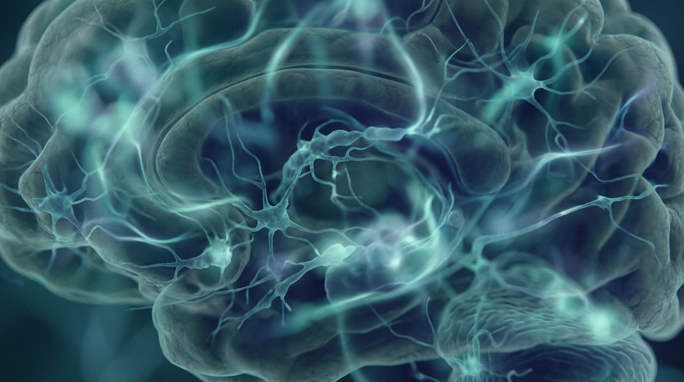
For most of the twentieth century, the hours before a migraine headache were noted but not fully characterized as part of the attack itself. The yawning, the food cravings, the mood change, the neck stiffness, the sensitivity to light and sound — these were called premonitory symptoms, a term that implies they are signals pointing toward a future event rather than a current one.
The neuroimaging evidence has progressively complicated that framing. A 2023 narrative review by Karsan and Goadsby, published in The Journal of Headache and Pain, examined imaging studies conducted during the premonitory phase — the period that begins up to 48 hours before headache onset. What those studies consistently showed was early dysfunction in three regions: the hypothalamus, the midbrain, and the limbic system.
These are not regions associated with headache in a simple or localized sense. They are regions involved in sensory processing, pain modulation, emotional state, and homeostatic regulation — the brain’s continuous management of its own internal equilibrium. Their disruption during the premonitory phase means that the migraine attack is not simply accumulating somewhere and waiting to release. The brain is already in a disrupted state. Pain thresholds are already shifting. Sensory filters are already changing.
Karsan and Goadsby also identified a clinical implication: calcitonin gene-related peptide — CGRP, a protein that plays a central role in migraine pain signaling — begins to be released during the premonitory phase. This matters because several established migraine treatments work by targeting CGRP or its receptor. Whether those treatments are more effective when taken during the premonitory window, rather than after headache onset, is an area of active investigation. The answer depends on understanding the premonitory phase as a real physiological event rather than a waiting room.
What the Hypothalamus Is Doing During Those Hours
Of all the brain regions implicated in the premonitory phase, the hypothalamus is the most studied. It is a small structure at the base of the brain, but its regulatory reach is extensive. It governs sleep-wake cycles, appetite, body temperature, hormonal release, and the coordination of the body’s stress response. It is, in a meaningful sense, the brain’s thermostat — and in the premonitory phase of a migraine, something about its calibration is off.
A 2022 paper by Gollion, De Icco, Dodick, and Ashina, published in The Journal of Headache and Pain, examined the case for hypothalamic dysfunction as the primary driver of the premonitory phase. Their analysis worked backward from the specific symptoms that characterize this period and traced each one to the neurotransmitter systems the hypothalamus controls.
Yawning, one of the most consistently reported premonitory symptoms, is modulated by dopaminergic pathways in the hypothalamus. The food cravings that many people with migraines experience before headache onset — specifically for carbohydrates and sweets — have been linked to neuropeptide Y signaling, which the hypothalamus uses to regulate appetite and energy balance. The fatigue and mood changes that characterize the early premonitory period connect to changes in orexin neurotransmission; orexins are hypothalamic signaling molecules that normally support wakefulness and stabilize arousal levels.
The picture that emerges from Gollion et al. is not a single malfunction in a single neurotransmitter system. It is a disruption that is running simultaneously across several systems, all coordinated by the same structure. This explains why the premonitory symptoms span physiology in the way they do — energy, appetite, arousal, mood, temperature sensitivity — without mapping to any single organ or any single chemical.
The paper also draws attention to the hypothalamus’s anatomical connections to the trigeminocervical complex — the region of the brainstem through which pain signals from the head travel. Hypothalamic activation during the premonitory phase does not only produce the symptoms we notice in the hours before a headache. It also directly modulates pain-related transmission in the pathway that will carry the headache pain itself. The premonitory phase and the pain phase are not separate events separated by time. They are connected at the level of the circuit, and the connection is already active before the headache arrives.
Neuroimaging has confirmed the hypothalamic activation that Gollion and colleagues describe. PET and fMRI studies examining people during the premonitory period — when they are aware that a migraine is coming but have not yet developed headache — have shown measurable increases in hypothalamic activity. The finding has been replicated across independent studies and multiple groups.
How the Pain System Gets Drawn In: The Trigeminovascular Pathway
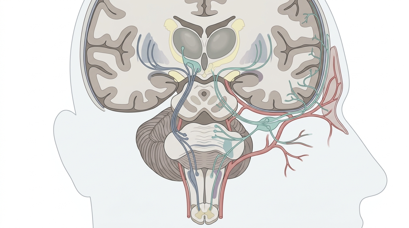
The pain of a migraine headache originates in a system called the trigeminovascular pathway: the network of blood vessels surrounding the brain, the trigeminal nerve endings that innervate those vessels, and the brainstem pathways through which their signals travel.
A 2025 paper by Christensen, Ashina, and Ashina, published in The Journal of Headache and Pain, examined what they describe as the “vessel-to-neuron” trigeminovascular hypothesis of migraine pathogenesis. Their analysis argues that intracranial vascular activation — changes in the meningeal blood vessels, the membranes surrounding the brain — triggers the trigeminal nerve endings embedded in those vessels to release a cascade of neuropeptides. The most prominent of these is CGRP.
CGRP is a potent vasodilator. When it is released from trigeminal nerve endings in the meninges, several things follow. It signals nearby mast cells to release inflammatory mediators. It causes local vasodilation in the meningeal vessels. And it sensitizes the nociceptors — the pain-sensing nerve endings — embedded in the meningeal tissue. This process, called neurogenic inflammation, creates a local environment in which the trigeminovascular nociceptors become progressively more sensitive to normally innocuous stimuli: normal blood pressure pulsation, gentle movement of the head, changes in body position.
This is the physiology behind the throbbing quality of migraine headache. It is not a new pain that arrives. It is that the normal rhythmic pulsation of the meningeal blood vessels — which happens continuously and without any sensation under ordinary circumstances — now crosses the pain threshold because the sensory system has been primed. The signal was always there; the threshold moved.
Christensen and colleagues present their paper explicitly as a “pro” argument in a contested area. The opposing framework proposes that neuronal changes precede vascular ones — that the nervous system’s activation drives the vascular changes rather than the other way around. They do not claim to have definitively resolved this debate; they acknowledge it is ongoing. What their synthesis establishes is that once this sequence is engaged, CGRP plays a central and consistent role throughout it, and that the pathway from vascular activation to trigeminal sensitization to central pain processing is biologically coherent and well-supported by the evidence they review.
Cortical Spreading Depression and the Aura Connection
Approximately one in three people who have migraines also experience aura: a wave of neurological symptoms that typically precedes the headache phase. Visual aura is the most common — a shimmering arc, a blind spot, geometric patterns that move across the visual field and resolve over 20 to 60 minutes. Some people experience sensory aura, a spreading numbness or tingling that moves across one side of the face or hand. A smaller number experience difficulties with language. The aura passes; the headache then begins.
The biological mechanism underlying this experience is a phenomenon called cortical spreading depression, or CSD. It is a slow wave of intense neuronal depolarization — a mass electrical discharge — followed by a period of suppressed neural activity, that propagates across the surface of the cortex at approximately 3 to 5 millimeters per minute. As the wave travels through the visual cortex at the back of the brain, it produces the characteristic visual disturbances. As it moves into somatosensory cortex, it produces the sensory symptoms. The wave is followed by reduced cortical activity, which corresponds to the negative symptoms — the blind spot, the numbness.
A 2025 paper by Charles and Goadsby, published in Headache: The Journal of Head and Face Pain, examines the cortical spreading depression–migraine aura hypothesis and argues, based on accumulated evidence, that CSD is the mechanism by which migraine aura is produced — not merely a correlate of it. Their review draws on animal models, electrophysiology, imaging studies in humans, and pharmacological evidence to make this case.
Critically, CSD is also connected to the pain cascade. The wave of depolarization activates the trigeminal nervous system and upregulates CGRP release. This means the cortical and trigeminovascular phases of a migraine attack are not independent tracks running in parallel. CSD links them: the cortical event feeds back into the pain signaling pathway that was already being primed by the hypothalamic changes of the premonitory phase.
A longstanding and still unresolved question is whether CSD also occurs in people who do not report aura symptoms. CSD propagating through cortical regions that do not produce detectable sensory or visual phenomena could in principle occur without any reported aura. Some researchers have proposed this as a mechanism for migraine without aura; the hypothesis remains under investigation. The absence of reported aura does not confirm the absence of cortical changes.
What the Research Does Not Yet Establish
The research described in this article represents a substantial advance in understanding migraine as a neurological condition. It is also important to state clearly what it does not establish.
The sequence described — premonitory hypothalamic dysfunction, trigeminovascular activation, CGRP cascade, and in some cases cortical spreading depression — is a model of how a migraine attack unfolds. Models are supported by population-level evidence; they describe patterns across studied groups. They do not specify the exact mechanism operating in every individual who has migraines, and migraine presentation varies considerably. Some people reliably experience a premonitory phase they can identify; others do not, or do not recognize it as such. The relationship between premonitory symptoms and subsequent headache onset is probabilistic, not invariable.
The causal direction of several steps in this cascade remains contested. Christensen and colleagues present the vessel-to-neuron model explicitly as one side of a live scientific argument. The competing neuronal-first view has its own body of supporting evidence. Charles and Goadsby offer what they characterize as definitive evidence for CSD as the mechanism of aura — but the precise relationship between CSD and non-aura migraine is not settled. These are active debates, not resolved questions dressed as settled ones.
The neuroimaging studies of the premonitory phase reviewed by Karsan and Goadsby have generally involved small sample sizes and methodological variation between studies. Consistency of findings across different imaging protocols, patient populations, and laboratories is ongoing work.
Finally, all of these findings are group-level results. They describe what researchers observed across cohorts of participants. The mechanisms described here are real — they reflect measured, reproducible phenomena at the studied level. What they predict about any specific individual’s migraine experience is a separate and substantially harder question, and the researchers are generally careful to say so.
Frequently Asked Questions
Is a migraine actually happening during the premonitory phase, before the headache starts?
Based on neuroimaging evidence reviewed by Karsan and Goadsby in 2023, the answer is yes. During the premonitory phase — which can begin up to 48 hours before headache onset — measurable dysfunction is already present in the hypothalamus, midbrain, and limbic regions involved in pain modulation and sensory processing. CGRP, a key neuropeptide in migraine pain signaling, begins to be released during this phase. The traditional framing of premonitory symptoms as signals pointing toward a future attack does not capture this: the neurological process is already underway.
Why do some people crave sweet or carbohydrate-heavy foods before a migraine headache?
Gollion and colleagues, in a 2022 analysis published in The Journal of Headache and Pain, traced this symptom to neuropeptide Y signaling in the hypothalamus. Neuropeptide Y is a hypothalamic transmitter that regulates appetite and energy balance. During the premonitory phase, hypothalamic dysfunction appears to alter this signaling in ways that drive food cravings, particularly for carbohydrates and sweets. The craving is not a behavioral habit or a simple sugar deficit. It reflects an altered neurochemical state in a brain structure that regulates energy across the whole body. Yawning — another common premonitory symptom — traces to dopaminergic pathways in the same region.
What is CGRP, and why does it come up so often in discussions of migraine?
CGRP stands for calcitonin gene-related peptide. It is a signaling molecule released by trigeminal nerve endings in the meningeal blood vessels surrounding the brain. When released, it causes local vasodilation, signals nearby cells to release inflammatory substances, and sensitizes the pain-sensing nerve endings in meningeal tissue. This process — called neurogenic inflammation — is central to how the pain of migraine is generated and maintained. CGRP is also released during the premonitory phase, before headache onset. A class of migraine treatments called gepants and a class of preventive therapies called anti-CGRP monoclonal antibodies both target this pathway.
Do all people who have migraines experience a premonitory phase?
Not all people who have migraines report identifiable premonitory symptoms, and the research does not establish that a full premonitory phase occurs in every migraine. What the neuroimaging evidence shows is that measurable brain changes precede headache onset in the people who have been studied — but studies of the premonitory phase have generally involved participants who were aware their migraines were coming. Whether the same hypothalamic and limbic changes occur in people who report no premonitory symptoms, and simply go unnoticed, or are absent in some migraine types, is a question the research has not settled.
Is migraine aura dangerous, and what is happening in the brain during it?
Migraine aura is caused by cortical spreading depression — a slow wave of intense neuronal depolarization followed by suppressed neural activity that moves across the surface of the cortex at roughly 3 to 5 millimeters per minute. As the wave travels through visual cortex, it produces the visual disturbances most people associate with aura. Charles and Goadsby, in a 2025 paper in Headache, presented what they describe as definitive evidence for this mechanism. In the great majority of people with migraines and aura, the aura itself is not dangerous — it is unpleasant and disruptive, but it reflects a transient cortical event. A small number of people have experienced stroke-like symptoms during aura; anyone experiencing new or prolonged aura symptoms, or aura without subsequent headache for the first time, should be evaluated by a physician.
Sources
- Karsan N, Goadsby PJ. Neuroimaging in the pre-ictal or premonitory phase of migraine: a narrative review (2023) — The Journal of Headache and Pain
- Gollion C, De Icco R, Dodick DW, Ashina H. The premonitory phase of migraine is due to hypothalamic dysfunction: revisiting the evidence (2022) — The Journal of Headache and Pain
- Christensen RH, Ashina H, Ashina M. The vessel-to-neuron trigeminovascular hypothesis of migraine pathogenesis — the 'pro' argument (2025) — The Journal of Headache and Pain
- Charles AC, Goadsby PJ. The cortical spreading depression/migraine aura hypothesis — Finally some definitive evidence (2025) — Headache: The Journal of Head and Face Pain / PubMed
Understand Your Nervous System
How the nervous system processes sensory information, regulates arousal, and responds to internal signals is at the center of what we work with at NeuroBalance. If you're curious about what one-on-one nervous system work looks like — whether you're dealing with migraine-related sensitivity, chronic stress, or other patterns — we offer a free initial consultation. Our practice is private, personal, and focused on your specific nervous system.
Schedule Your Free Consultation
Call us: (424) 625-5445 · Los Angeles, CA
Disclaimer: This content is for informational purposes only and does not constitute medical advice, diagnosis, or treatment. LENS Neurofeedback is not FDA-approved for all conditions mentioned. Please consult with a qualified healthcare provider before beginning any new treatment program.

A balanced brain is a better brain for a happier life